Page 43 - IJB-2-1
P. 43

Christopher Chi Wai Tse, Shea Shin Ng, Jonathan Stringer, et al.

                                                               D). Figure 5 show cells aligning to the same orienta-
                                                               tion  as the channel, and through the use of ImageJ
                                                               software, the orientation of cells could be clearly ob-
                                                               served following the direction of the channel. Cell
                                                               alignment could be investigated with this methodolo-
                                                                                                      [8]
                                                               gy as in the research reported by Duclos et al. , who
                                                               described how  NIH-3T3  mouse embryo fibroblasts
                                                               aligned on confined strips from 30 µm to 1.5 mm.
                                                                 After fibroblasts or Schwann cells were seeded,
                                                               they  adhered, had  spread  and  thereafter proliferated
                                                               into the  desired  positions and orientation.  The  wax
                                                               structures were easily removed from the samples with
                                                               a sharp scalpel, which was used to physically peel the
                                                               wax off, to leave the cells to grow without any space
                                                               limitations.  Figure 6  shows images of the fabricated
                                                               wax  structures, patterned  cells before and  after wax
                                                               removal. Cells maintained their position and orienta-
                                                               tion for at least 7 days after seeding as shown.

                                                               3.3 Cell Proliferation After Removal of Wax
                                                               A wax scaffold with channel widths of 40 µm and 30
                                                               µm  connected together  at one  end  were created  and
                                                               fibroblasts were seeded into the structure and cultured.
                                                               After 2 days of culture and prior to wax removal, fi-
                                                               broblasts had aligned  within  the  shape  of  the  open
                                                               channels (Figure 7A). Upon wax removal, cells were
                                                               not limited to the channel space (Figure 7B) and had
                                                               spread to cover the substrate, which was observed 24
                                                               to 48 hours after wax removal (Figure 7C and D). The
                                                               wax  printing  technique  could  therefore  be  used  in-
                                                               itially to  deposit cells in the desired  areas spatially.
                                                               The ability  of the wax to be removed from the sub-
                                                               strate, without the addition of new substances into the
                                                               environment, allowed  for  a  new  method  to  analyse
                                                               cell  migration and proliferation on open substrates
                                                               to be studied over time. We observed a high level of
                                                               alignment to the direction of channels with diameters
                                                               of less than 160 µm, and consequently the authors of
                                                               this paper suggest that this work would have imme-
                                                               diate applications in cell migration and  “scratch as-
                                                               say”  type studies, commonly used in wound healing

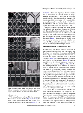
            Figure 3. Micrographs of printed wax on glass.  (A–C) show   and cancer cell migration research.
            different patterns that  were produced.  Magnified  light  micro-  Cells were able to  remain  attached  onto the sub-
            scopy images are shown in the small frame. Bar = 200 µm.   strate even after wax removal. An exception to this
                                                               occurred when cell concentration exceeded confluence.
            cells to grow.                                     From this study, when cell density began to reach con-
                                                                                                     2
               With  isolated  arrays, cells proliferated  in  random   fluency  (typically  more than 300 cells/mm ) an  in-
            shapes with no obvious direction (Figure 4A and B), but   creased proportion of cells detached. All cells within
            when  a linear patterned  structure  was  created, cells   the scaffold were removed when cell density reached
                                                                                2
            aligned in the direction of the structure (Figure 4C and   above 700 cells/mm . It was postulated that this was
                                        International Journal of Bioprinting (2016)–Volume 2, Issue 1      39
   38   39   40   41   42   43   44   45   46   47   48