
Page 73 - IJB-2-1
P. 73
Hui Wang, Sanjairaj Vijayavenkataraman, Yang Wu, et al.
Figure 9. SEM Images of semi-lunar PCL scaffolds.
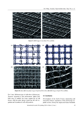
Figure 10. (A) & (B) Conceptual images of spiral scaffold. (C) & (D) SEM Images of spiral PCL scaffolds.
D = 3 mm. Different types of cells show different pro-
liferation responses to the environment. Spiral scaf- 4. Conclusion
folds may be suitable for tissue engineering of soft Development of the field of tissue engineering and
tissues, with the spiral design improving the cell mi- successful clinical translation of this technology de-
gration and formation of cell-cell junctions. pends on how closely the engineered tissue biomimic
International Journal of Bioprinting (2016)–Volume 2, Issue 1 69