
Page 77 - IJB-2-1
P. 77
Ryan D. Boehm, Panupong Jaipan, Kai-Hung Yang, et al
normal cooking temperatures does not necessarily 2. Materials and Methods
[1]
alter the histamine levels within the fish .
Due to concerns associated with histamine conta- 2.1 Microneedle and Lateral Flow Test Holder
mination of fish, a number of methods have been de- To sample the tuna flesh for histamine, arrays of mi-
veloped to screen fish flesh and ensure that it does not croneedles were used to capture fluid from the tuna
contain dangerous levels of histamine. Screening of samples. A custom lateral flow test strip holder was
histamine levels in fish may be conducted using a va- designed to stabilize a microneedle array, allowing the
riety of methods, including high purity liquid chro- sampled fluid to be washed off of the microneedle
matography (HPLC) [11] , enzymatic test kits, enzyme- array and into a reservoir for wetting of a lateral flow
linked immunosorbent assays (ELISA), and lateral test (Figure 1). Both of these components were custom
flow immunochromatographic test strips [12] . The later- designed using computer-aided design software (Solid-
al flow test strips are particularly useful for fieldwork Works Education Edition 2014–2015, Dassault Systémes
since they do not require complex equipment for SolidWorks Corporation, Concord, NH, USA). The
analysis. Lateral flow tests, sometimes referred to as microneedle arrays were composed of nine offset mi-
dipsticks, are used in many environmental and health- croneedles, which exhibited a thin pyramidal shape
care applications (e.g., colorimetric pregnancy tests) [13] . and a trapezoidal eyelet design to capture fluid (Figure
For example, lateral flow tests have been used for bo- 1A). The test strip and microneedle holder (Figure 1C)
tulinum neurotoxin, aflatoxin B1, and virus detec- was designed with three chambers: (i) a washed sam-
®
tion [14–17] . One such lateral flow test is the Reveal for ple reservoir for placement of the lateral flow test, (ii)
®
Histamine test kit (Neogen Corporation, Lansing, MI, the microneedle holder/wash chamber, and (iii) an
USA), which can screen for histamine in tuna and
®
mahi-mahi; the Reveal for Histamine test kit was
used as the histamine detection mechanism in this
study.
One procedure that is described in many histamine
detection methods is homogenization of fish flesh;
most assays are performed on fluid extracts from the
homogenized fish flesh. In this study, we investigated
use of a microstereolithography-prepared microneedle
sampling system for detecting histamine in fish flesh.
Systems containing microneedle arrays have previ-
ously been developed for sampling of analytes in tran-
sdermal blood and/or interstitial fluid [18–20] . For in-
stance, systems containing microneedles arrays have
been developed for detection of glutamate [21] , glu-
cose [22,23] , and potassium ions [24] . In this study, micro-
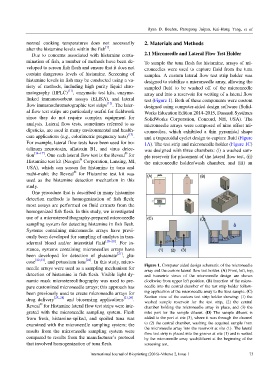
needle arrays were used as a sampling mechanism for Figure 1. Computer aided design schematic of the microneedle
array and the custom lateral flow test holder. (A) Front, left, top,
detection of histamine in fish flesh. Visible light dy- and isometric views of the microneedle design are shown
namic mask microstereolithography was used to pre- clockwise from upper left position. (B) Insertion of the micro-
pare customized microneedle arrays; this approach has needle into the central chamber of the test strip holder follow-
been previously used to create microneedle arrays for ing application of the microneedle array to the tuna sample. (C)
drug delivery [25–28] and biosensing applications [21,29] . Section view of the custom test strip holder showing: (1) the
washed sample reservoir for the test strip, (2) the central
®
Reveal for Histamine lateral flow test strips were inte- chamber holding the microneedle array in place, and (3) the
grated with the microneedle sampling system. Flesh inlet port for the sample diluent. (D) The sample diluent is
from fresh, histamine-spiked, and spoiled tuna was added to the port at site (3), where it runs through the channel
examined with the microneedle sampling system; the to (2) the central chamber, washing the acquired sample from
results from the microneedle sampling system were the microneedle array into the reservoir at site (1). The lateral
flow test strip is placed into the groove at site (1) and is wetted
compared to results from the manufacturer’s protocol by the microneedle array wash/diluent at the beginning of the
that involved homogenization of tuna flesh. screening test.
International Journal of Bioprinting (2016)–Volume 2, Issue 1 73