Page 240 - IJB-10-2
P. 240
International Journal of Bioprinting                       G40T60@WNT5A promotes osteoblast differentiation


            the sample was heated for 3 min using a sputter coating   (ab150165, Abcam, UK). After incubation at 4°C for 30
            device (SCD 005; BAL-TEC, Liechtenstein) at a current of   min, unbound antibodies were washed away with PBS.
            30 mA. 52                                          The cells successfully targeted by the labeled antibodies
                                                               were analyzed by means of flow cytometry. To study the
            2.13. Mechanical experiment                        differentiation of BMSCs into osteoblasts, adipocytes, and
            The G40T60 bracket was placed on a load cell with a   chondrocytes, an array of approaches including reagent
            capacity of 500 N on a universal testing machine (EZ   kit (PD-003/4/5, Procell), Alizarin red staining (ARS),
            Test; SHIMADZU, Japan). The compressive strength of   oil red O staining, and Alcian blue staining were used to
            the bracket was measured at a crosshead speed of 5 mm/  observe  the  osteogenic,  adipogenic,  and  chondrogenic
            min. The compressive strength of five samples for each   differentiation abilities of BMSCs. The above methods
            proportion was determined for the measurements of mean   confirmed the osteogenic, adipogenic, and chondrogenic
            and standard deviation. 52                         differentiation capacity of the BMSCs used in this study
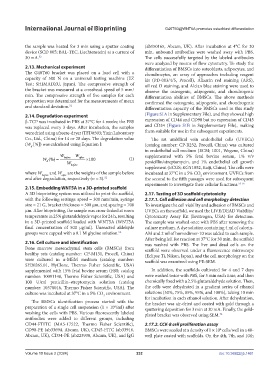
            2.14. Degradation experiment                       (Figure S1A in Supplementary File), and they showed high
            β-TCP was incubated in PBS at 37°C for 4 weeks; the PBS   expression of CD44 and CD90 but no expression of CD45
            was replaced every 3 days. After incubation, the samples   and CD34 (Figure S1B in Supplementary File), making
            were dried using a freeze-dryer (TFD8503; Yixin Laboratory   them suitable for use in the subsequent experiments.
            Co., Ltd., China) for 14 or 28 days. The degradation value   The rat umbilical vein endothelial cells (UVECs)
            (W  [%]) was calculated using Equation I:          (catalog number: CP-R232, Procell, China) was cultured
              d
                                                               in endothelial cell medium (ECM; 1001, Wegene, China)
                           W     W                            supplemented with 5% fetal bovine serum, 1% v/v
                   W (%)   before  after  100         (I)    penicillin/streptomycin, and 1% endothelial cell growth
                     d
                              W
                               before
                                                               supplement (ECGS; KGY1052, Kaiji, China). The cells were
            where W before  and W  are the weights of the sample before   incubated at 37°C in a 5% CO  environment. UVECs from
                           after
                                                                                       2
            and after degradation, respectively (n = 3). 52    the second to the fifth passages were used for subsequent
                                                               experiments to investigate their cellular functions. 53-55
            2.15. Embedding WNT5A in a 3D-printed scaffold
            A 3D bioprinting system was utilized to print the scaffold,   2.17. Testing of 3D scaffold cytotoxicity
            with the following settings: speed = 300 mm/min, syringe   2.17.1. Cell adhesion and cell morphology detection
            size = 21 G, bracket thickness = 500 µm, and spacing = 700   To investigate the cell viability and adhesion of BMSCs and
            µm. After bioprinting, the sample was cross-linked at room   UVECs on the scaffold, we used the LIVE/DEAD Viability/
            temperature in 25% glutaraldehyde vapor for 24 h, resulting   Cytotoxicity Assay Kit (Invitrogen, USA) for detection.
            in a 3D-printed scaffold loaded with WNT5A (WNT5A   The sample was washed once with PBS after removing the
            final concentration of 500 μg/ml). Unreacted aldehyde   culture medium. A dye solution containing 4 ml of calcein-
            groups were capped with a 0.1 M glycine solution. 52  AM and 2 ml of homodimer-10 was added to each sample.
                                                               After being left for reaction at 37°C for 30 min, the scaffold
            2.16. Cell culture and identification              was washed with PBS. The live and dead cells on the
            Bone marrow mesenchymal stem cells (BMSCs) from    scaffold were observed under a fluorescence microscope
            healthy rats (catalog number: CP-M131, Procell, China)   (Eclipse Ti, Nikon, Japan), and the cell morphology on the
            were cultured in α-MEM medium (catalog number:     scaffold was examined using FE-SEM.
            SH30265.01, HyClone, Thermo Fisher Scientific, USA)
            supplemented with 15% fetal bovine serum (FBS; catalog   In addition, the scaffolds cultivated for 4 and 7 days
            number: 10091148, Thermo Fisher Scientific, USA) and   were washed twice with PBS, for 5 min each time, and then
            100 U/ml penicillin–streptomycin solution (catalog   chemically fixed with a 2.5% glutaraldehyde solution. Then,
            number: 10378016, Thermo Fisher Scientific, USA). The   the cells were dehydrated in a gradient series of ethanol
            culture was incubated at 37°C in a 5% CO  environment.  solutions (50%, 75%, 85%, 95%, and 100%), taking 10 min
                                             2
               The BMSCs identification process started with the   for incubation in each ethanol solution. After dehydration,
                                                               the bracket was air-dried and coated with gold through a
            preparation of a single cell suspension (1 × 10 /ml) after   sputtering deposition for 3 min at 30 mA. Finally, the gold-
                                                  6
            washing the cells with PBS. Various fluorescently labeled   plated bracket was observed using SEM. 52
            antibodies  were added to  different  groups,  including
            CD44-FTITC (MA5-17522, Thermo Fisher Scientific),   2.17.2. CCK-8 cell proliferation assay
            CD90-PE (ab33694, Abcam, UK), CD45-FITC (ab33916,   BMSCs were seeded at a density of 1 × 10  cells/well in a 48-
                                                                                               4
            Abcam, UK), CD34-PE (ab223930, Abcam, UK), and IgG   well plate coated with scaffolds. On the 4th, 7th, and 10th


            Volume 10 Issue 2 (2024)                       232                                doi: 10.36922/ijb.1461

