
Page 250 - IJB-10-2
P. 250
International Journal of Bioprinting G40T60@WNT5A promotes osteoblast differentiation
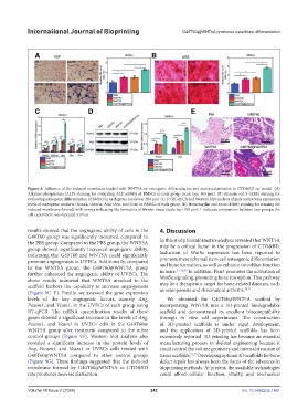
Figure 8. Influence of the induced membrane loaded with WNT5A on osteogenic differentiation and neovascularization in CTO&BD rat model (A)
Alkaline phosphatase (ALP) staining for evaluating ALP activity of BMSCs in each group (scale bar: 400 μm). (B) Alizarin red S (ARS) staining for
evaluating osteogenic differentiation of BMSCs in each group (scale bar: 200 μm). (C, D) RT-qPCR and Western blot analysis of gene and protein expression
levels of osteogenic markers (Runx2, Osterix, Alpl, Opn, and Ocn) in BMSCs of each group. (E) Hematoxylin and eosin (H&E) staining for staining the
induced membrane formed, with arrows indicating the formation of fibrous tissue (scale bar: 100 μm). * indicates comparison between two groups; the
cell experiment was repeated 3 times.
results showed that the angiogenic ability of cells in the 4. Discussion
G40T60 group was significantly increased compared to
the PBS group. Compared to the PBS group, the WNT5A In this study, bioinformatics analysis revealed that WNT5A
group showed significantly increased angiogenic ability, may be a critical factor in the progression of CTO&BD.
indicating that G40T60 and WNT5A could significantly Induction of Wnt5a expression has been reported to
promote angiogenesis in UVECs. Additionally, compared promote mesenchymal stem cell osteogenic differentiation
to the WNT5A group, the G40T60@WNT5A group and bone formation, as well as enhance osteoblast function
33-36,69
further enhanced the angiogenic ability of UVECs. The in mice. In addition, Pkn3 promotes the activation of
above results indicated that WNT5A attached to the Wnt5a signaling, promoting bone resorption. This pathway
scaffold harbors the capability to increase angiogenesis may be a therapeutic target for bone-related diseases, such
70,71
(Figure 9C–E). Finally, we assessed the gene expression as osteoporosis and rheumatoid arthritis.
levels of the key angiogenic factors, namely Ang, We obtained the G40T60@WNT5A scaffold by
Pecam1, and Vcam1, in the UVECs of each group using incorporating WNT5A into a 3D-printed biodegradable
RT-qPCR. The mRNA quantification results of these scaffold and demonstrated its excellent biocompatibility
genes showed a significant increase in the levels of Ang, through in vitro cell experiments. The construction
Pecam1, and Vcam1 in UVECs cells in the G40T60@ of 3D-printed scaffolds is under rapid development,
WNT5A group after treatment compared to the other and the application of 3D-printed scaffolds has been
control groups (Figure 9F). Western blot analysis also extensively reported. 3D printing has become an essential
revealed a significant increase in the protein levels of manufacturing process in skeletal engineering because it
Ang, Pecam1, and Vcam1 in UVECs cells treated with could control the volume geometry and internal structure of
G40T60@WNT5A compared to other control groups tissue scaffolds. 72,73 Developing optimal 3D scaffolds for bone
(Figure 9G). These findings suggested that the induced defect repair has always been the focus of the advances in
membrane formed by G40T60@WNT5A in CTO&BD bioprinting methods. At present, the available technologies
rats promotes neovascularization. could afford cellular function, vitality, and mechanical
Volume 10 Issue 2 (2024) 242 doi: 10.36922/ijb.1461