
Page 400 - IJB-10-2
P. 400
International Journal of Bioprinting Automated bioink mixer improves bioprinting
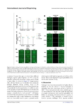
Figure 8. Machine mixing of bioinks using different cell types and hydrogels. (A) Metabolic activity of different cell types after automated mixing with 6%
alginate as evaluated by XTT assays 1 day after bioprinting. The values were normalized to the respective average for each cell type. Cell viability determined
by live/dead staining. Cells were treated with calcein AM and ethidium homodimer-1 to stain living cells green and dead cells red. (C) Metabolic activity
of HEK293-GFP cells in different hydrogels (alginate, alginate/gelatin, and PAA). The values were normalized to the respective average for each cell type.
(D) Cell distribution as determined by fluorescence microscopy directly after mixing. n = 3; data are expressed as mean ± SD.
constructs. To assess this aspect, we tested three different which requires both high homogeneity and stiffness of the
hydrogels: alginate, gelatin/alginate, and PAA, using the bioink. These results highlight the good printability of the
regenHU 3DDiscovery bioprinter to print various models bioinks after machine mixing.
on glass slides. As shown in Figure S7 (Supplementary
File), all three bioinks can be extruded into continuous 4. Discussion
hydrogel filaments during printing, which is the Hydrogels have been demonstrated to be highly effective
fundamental requirement for extrusion-based bioprinting. matrices for 3D cell culture. 34-36 Viscous hydrogels, in
We specifically tested the production of a waffle structure, particular, have been extensively utilized as bioinks in
as it is a popular model in 3D bioprinting research due to its EBB. 37,38 These hydrogels not only create a suitable aqueous
simple and stable architecture, as well as its porosity which 3D environment for cells, but also enable the fabrication
ensures a desirable level of cell survival. 29-33 All bioinks of complex architectures with excellent printability and
mixed with the automated device were found to be suitable fidelity by preventing deformation driven by gravity or
for printing the waffle model. Furthermore, the bioinks surface tension. The preparation of viscous bioinks,
7,39
were also successfully used to print a pyramid model, however, presents challenges. While vigorous methods
Volume 10 Issue 2 (2024) 392 doi: 10.36922/ijb.1974