
Page 458 - IJB-10-2
P. 458
International Journal of Bioprinting 3D-printed bioceramic scaffolds for bone regeneration
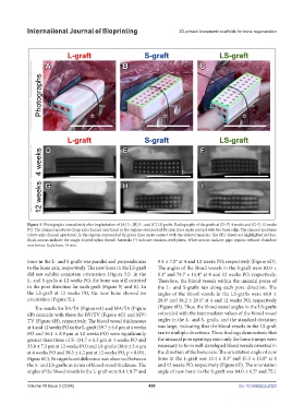
Figure 3. Photographs immediately after implantation of (A) L-, (B) S-, and (C) LS-grafts. Radiographs of the grafts at (D–F) 4 weeks and (G–I) 12 weeks
PO. The channel apertures (long-axis channel apertures) in the regions surrounded by cyan lines make contact with the bone edge. The channel apertures
(short-axis channel apertures) in the regions surrounded by green lines make contact with the skeletal muscles. The PET sheets are highlighted in blue.
Black arrows indicate the single-looped nylon thread. Asterisks (*) indicate stainless-steel plates. White arrows indicate gaps regions without abundant
new bones. Scale bars: 10 mm.
bone in the L- and S-grafts was parallel and perpendicular 9.4 ± 7.0° at 4 and 12 weeks PO, respectively (Figure 6D).
to the bone axis, respectively. The new bone in the LS-graft The angles of the blood vessels in the S-graft were 82.0 ±
did not exhibit consistent orientation (Figure 5I). In the 8.1° and 74.7 ± 11.4° at 4 and 12 weeks PO, respectively.
L- and S-grafts at 12 weeks PO, the bone was still oriented Therefore, the blood vessels within the uniaxial pores of
in the pore direction for each graft (Figure 5J and K). In the L- and S-grafts ran along each pore direction. The
the LS-graft at 12 weeks PO, the new bone showed no angles of the blood vessels in the LS-grafts were 46.8 ±
orientation (Figure 5L). 28.6° and 38.2 ± 29.1° at 4 and 12 weeks PO, respectively
The results for BA/TA (Figure 6A) and MA/TA (Figure (Figure 6D). Thus, the blood vessel angles in the LS-grafts
6B) coincide with those for BV/TV (Figure 4G) and MV/ coincided with the intermediate values of the blood vessel
TV (Figure 4H), respectively. The blood vessel thicknesses angles in the L- and S- grafts, and the standard deviation
at 4 and 12 weeks PO in the L-graft (59.7 ± 6.0 µm at 4 weeks was large, indicating that the blood vessels in the LS-graft
PO and 56.1 ± 8.9 µm at 12 weeks PO) were significantly ran in multiple directions. These findings demonstrate that
greater than those of S- (34.7 ± 6.3 µm at 4 weeks PO and the uniaxial pore openings onto only the bone stumps were
33.8 ± 7.3 µm at 12 weeks PO) and LS-grafts (38.6 ± 3.4 µm necessary to form well-developed blood vessels oriented in
at 4 weeks PO and 38.3 ± 4.2 µm at 12 weeks PO, p > 0.001, the direction of the bone axis. The orientation angle of new
Figure 6C). No significant difference was observed between bone in the L-graft was 10.4 ± 8.3° and 15.3 ± 15.8° at 4
the S- and LS-grafts in terms of blood vessel thickness. The and 12 weeks PO, respectively (Figure 6E). The orientation
angles of the blood vessels in the L-graft were 8.4 ± 8.7° and angle of new bone in the S-graft was 84.0 ± 6.5° and 75.1
Volume 10 Issue 2 (2024) 450 doi: 10.36922/ijb.2323