
Page 460 - IJB-10-2
P. 460
International Journal of Bioprinting 3D-printed bioceramic scaffolds for bone regeneration
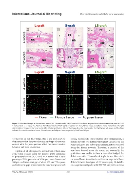
Figure 5. HE-stained images in the new bone area at (A–C) 4 weeks and (D–F) 12 weeks PO. Analyzed images of bone orientations within pores at (G–I)
4 weeks and (J–L) 12 weeks PO. Asterisks (*) indicate material. NB, FT, and AT denote new bone, fibrous tissue, and adipose tissue, respectively. The right
and left sides of images are the bone stump sides. The top and bottom sides of the images show the muscle sides. The highlighted red, green, and blue lines
indicate the orientations of new bones, fibrous tissues, and adipose tissue, respectively. Scale bars: 100 µm.
To the best of our knowledge, this is the first study to tissues, respectively. Three months after implantation, a
demonstrate that the pore direction and type of tissue in fibrous network was formed throughout the graft via the
contact with the pore aperture affect the tissue invasion pores and gaps, and subsequent mineralization occurred
behavior and bone orientation. along the fibrous network. Therefore, a portion of the
Cipitria et al. attempted to reconstruct critical-sized new bone formed across the struts, and eventually, the
ovine tibial defects using 3D porous grafts composed graft alone was unable to form a bone that bridged the
of polycaprolactone (PCL) and TCP, which had a total defect, even after 12 months of implantation. Hara et al.
porosity of 70%, pore size of 1200 µm, strut diameter of compared bone formation in rat femoral segmental bone
300 µm, and inter-strut gap of about 100 µm. The pores defects between two types of 3D porous poly (L-lactide-
10
and inter-strut gaps opened onto the bone stumps and soft co-ε-caprolactone) grafts with 300−500 μm pores: one was
Volume 10 Issue 2 (2024) 452 doi: 10.36922/ijb.2323