
Page 459 - IJB-10-2
P. 459
International Journal of Bioprinting 3D-printed bioceramic scaffolds for bone regeneration
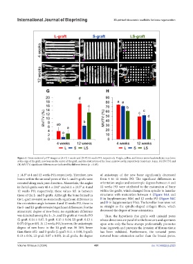
Figure 4. Cross sectional µ-CT images at (A–C) 4 weeks and (D–F) 12 weeks PO, respectively. Purple, yellow, and brown arrowheads indicate new bone
at the edge of the graft, new bone in the center of the graft, and the obstruction of the bone marrow cavity, respectively. Scale bars: 1 mm. (G) BV/TV, and
(H) MV/TV; significant differences are indicated by different letters (p < 0.05).
± 14.8° at 4 and 12 weeks PO, respectively. Therefore, new of anisotropy of the new bone significantly decreased
bones within the uniaxial pores of the L- and S-grafts were from 4 to 12 weeks PO. The significant differences in
oriented along each pore direction. Meanwhile, the angles orientation angles and anisotropic degrees between 4 and
in the LS-grafts were 48.4 ± 29.0° and 61.1 ± 23.5° at 4 and 12 weeks PO were attributed to the maturation of bone
12 weeks PO, respectively; these values fell in between within the grafts, which changed from spindle to lamellar
those of the L- and S-grafts. Although the bone formed in structures with maturation between 4 (Figure S4A and
the L-graft revealed no statistically significant difference in B in Supplementary File) and 12 weeks PO (Figure S4C
the orientation angle between 4 and 12 weeks PO, those in and D in Supplementary File). The lamellar lines were not
the S- and LS-grafts revealed significant differences. For the as straight as the spindle-shaped collagen fibers, which
anisotropic degree of new bone, no significant difference decreased the degree of bone orientation.
was detected among the L-, S-, and LS-grafts at 4 weeks PO Thus, the hypothesis that grafts with uniaxial pores
(L-graft: 0.14 ± 0.07, S-graft: 0.15 ± 0.04, LS-graft: 0.13 ± whose directions are parallel to the bone axis and apertures
0.07) (Figure 6F). At 12 weeks PO, however, the anisotropic open onto only the bone stumps preferentially promotes
degree of new bone in the LS-graft was 30–36% lower bone ingrowth and prevents the invasion of fibrous tissue
than those of L- and S-grafts (L-graft: 0.11 ± 0.08, S-graft: has been validated. Furthermore, the uniaxial pores
0.10 ± 0.06, LS-graft: 0.07 ± 0.05). In all grafts, the degree restored bone orientation earlier than the biaxial pores.
Volume 10 Issue 2 (2024) 451 doi: 10.36922/ijb.2323