
Page 461 - IJB-10-2
P. 461
International Journal of Bioprinting 3D-printed bioceramic scaffolds for bone regeneration
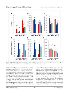
Figure 6. (A) BA/TA, (B) MA/TA, (C) blood vessel thickness, (D) angles of blood vessels relative to the pore direction, (E) orientation angles of new bones,
and (D) anisotropic degrees of new bones. Significant differences are indicated by different letters (p < 0.05).
tubular grafts with pores on the wall (66.3% of porosity), axis, into rabbit tibial segmental bone defects. The β-TCP
and the other was cylindrical grafts with 0/90 lay-down graft with multi-tubular pores promoted bone formation
36
pattern (53.1% of porosity). Although implantation of and restored bone bending strength faster than that with
37
these grafts promoted bone formation better compared to single-tubular pore. These previous findings support our
non-implantation of grafts, these grafts did not form bone finding that uniaxial pores should open only onto the bone
to bridge the defects even at 15 weeks PO. Conversely, Feng stumps to effectively reconstruct segmental bone defects.
et al. reported that one-dimensional porous β-TCP grafts Conventionally, the high porosity of the graft contributes
with a tubular pore opening only onto the bone stamps to bone formation while reducing the mechanical
promoted bone ingrowth in rabbit radius segmental strength. Thus, merely adjusting porosity is not sufficient
38
bone defects. Furthermore, Pan et al. implanted one- to achieve graft strength and bone formation. However,
24
dimensional porous β-TCP graft with single tubular pore in this study, we overcame this challenge: Although the
and multi-tubular pores, which were parallel to the bone total porosity of the L-graft was lower than that of the
Volume 10 Issue 2 (2024) 453 doi: 10.36922/ijb.2323