
Page 556 - IJB-10-2
P. 556
International Journal of Bioprinting OLS design for distal femur osseointegration
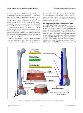
it is commonly assumed that the loading on the femur is the surrounding bones. The goal was to identify lattice
approximately four times the body weight. A downward structures that induced a strain close to, but not exceeding,
37
force of 2800 N was applied to the femoral head in the 4000 μ, indicating that the OLS exhibits osteoconductive
analyzed model, considering a patient’s body weight to properties and accelerates bone growth, thereby enhancing
be 70 kg. Fixed support was provided on the lateral and the osseointegration effect.
medial condyle surfaces of the distal femur. The contact
settings between the components were set to “frictional” 2.5. Biomechanical testing of implants and bone
to govern the interactions among the bone and screw, plate cement for distal femur reconstruction
and screw, as well as the lattice and bone. This frictional Biomechanical tests were conducted to assess the efficacy
setting incorporated the sliding resistance based on the of lattice structure on the distal femur reconstruction
friction coefficient, allowing these components to move implant in stimulating bone growth and enabling the
production of appropriate strain in the surrounding area.
relative to each other. This simulation represented the state These tests were conducted to investigate the differences
of the implant and bone before osseointegration occurred in surrounding bone growth (osteoconductive) among an
following surgery (Figure 3).
implant with OLS (OLS implant), an implant without lattice
During the analysis, different lattice structures structure (NLS implant), and bone cement (Radiopaque
were substituted for the lattice layers on the implant to Bone Cement, Stryker Corporation, Michigan, USA). The
assess the compression of the third principal strain on OLS implant and the NLS implant were manufactured
Figure 2. Overall model for distal femur defect reconstruction (modeling for finite element analysis).
Volume 10 Issue 2 (2024) 548 doi: 10.36922/ijb.2590