
Page 558 - IJB-10-2
P. 558
International Journal of Bioprinting OLS design for distal femur osseointegration
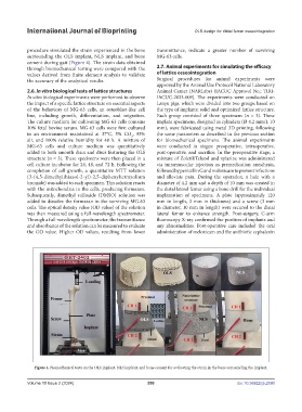
procedure simulated the strain experienced in the bone transmittance, indicate a greater number of surviving
surrounding the OLS implant, NLS implant, and bone MG-63 cells.
cement during gait (Figure 4). The strain data obtained
through biomechanical testing were compared with the 2.7. Animal experiments for simulating the efficacy
values derived from finite element analysis to validate of lattice osseointegration
the accuracy of the analytical results. Surgical procedures for animal experiments were
approved by the Animal Use Protocol National Laboratory
2.6. In vitro biological tests of lattice structures Animal Center (NARLabs) (IACUC Approval No.: TIRI-
In vitro biological experiments were performed to observe IACUC-2023-009). The experiments were conducted on
the impact of a specific lattice structure on essential aspects Lanyu pigs, which were divided into two groups based on
of the behaviors of MG-63 cells, an osteoblast-like cell the type of implants: solid and optimized lattice structure.
line, including growth, differentiation, and migration. Each group consisted of three specimens (n = 3). These
The culture medium for cultivating MG-63 cells contains implant specimens, designed as cylinders (Ø 4.2 mm/L 10
10% fetal bovine serum. MG-63 cells were first cultured mm), were fabricated using metal 3D printing, following
in an environment maintained at 37°C, 5% CO , 95% the same parameters as described in the previous section
2
air, and 100% relative humidity for 48 h. A mixture of for biomechanical specimens. The animal experiments
MG-63 cells and culture medium was quantitatively were conducted in stages: preoperative, intraoperative,
added to both smooth discs and discs featuring the OLS post-operative, and sacrifice. In the preoperative stage, a
structure (n = 3). These specimens were then placed in a mixture of Zoletil/Telazol and xylazine was administered
cell culture incubator for 24, 48, and 72 h. Following the via intramuscular injection as premedication anesthesia,
completion of cell growth, a quantitative MTT solution followed by penicillin G and meloxicam to prevent infections
(3-(4,5-dimethylthiazol-2-yl)-2,5-diphenyltetrazolium and alleviate pain. During the operation, a hole with a
bromide) was added to each specimen. This solution reacts diameter of 4.2 mm and a depth of 10 mm was created in
with the mitochondria in the cells, producing formazan. the distal lateral femur using a bone drill for the individual
Subsequently, dimethyl sulfoxide (DMSO) solution was implantation of specimens. A plate (approximately 120
added to dissolve the formazan in the surviving MG-63 mm in length, 3 mm in thickness) and a screw (3 mm
cells. The optical density value (OD value) of the solution in diameter, 10 mm in length) were secured to the distal
was then measured using a full-wavelength spectrometer. lateral femur to enhance strength. Post-surgery, C-arm
Through a full-wavelength spectrometer, the transmittance fluoroscopy X-ray confirmed the position of implants and
and absorbance of the solution can be measured to evaluate any abnormalities. Post-operative care included the oral
the OD value. Higher OD values, resulting from lower administration of meloxicam and the antibiotic cephalexin
Figure 4. Biomechanical tests on the OLS implant, NLS implant, and bone cement for evaluating the strain in the bone surrounding the implant.
Volume 10 Issue 2 (2024) 550 doi: 10.36922/ijb.2590