
Page 562 - IJB-10-2
P. 562
International Journal of Bioprinting OLS design for distal femur osseointegration
the efficacy of the lattice design in guiding bone growth of loading. While walking, the hip bears an average load
and achieving favorable osseointegration. Conversely, the of 238% of body weight. In a sitting position, the lumbar
46
solid implants, characterized by the non-porous structure, spine undergoes a pressure load of approximately 1698
47
restricted immature bone growth to the external periphery N, contrasting with 1076 N in a standing position.
of the implant, impeding penetration into the implant itself. Throughout the gait cycle, the tibial and ankle regions
This limitation significantly compromised the implant’s can bear loads up to 3.5 times the body weight. The
48
ability to integrate with the surrounding bone. Notably, the craniofacial region frequently experiences masticatory
29
volume of immature bone within the OLS surpassed the forces, with molars bearing a load of around 250 N.
original implant cavity volume. This observation suggests Comparing the loads in each area reveals significant
that immature bone smoothly grows not only into the differences, emphasizing the necessity to design implant
lattice structure but also along the implant’s periphery. lattices differently to accommodate various loading
The immature bone effectively encompasses the implant, conditions and expedite implant osseointegration.
thereby augmenting the implant’s osseointegration 4.2. Correlation between lattice structure
capability (Table 4). parameters and material properties
From a microcosmic perspective, it is essential for the lattice
4. Discussion structure on the implant surface to possess an appropriate
4.1. Optimal lattice structure design for implants in elastic modulus and favorable osteoconductive properties
distal femur mechanical conditions to facilitate bone growth into it. A crucial design criterion
From a macroscopic perspective, it is crucial to examine for distal femur defect reconstruction implant is selecting
the implant’s structure to ensure its capability to withstand lattice structure parameters whose elastic modulus aligned
10
the body’s weight and the daily-life force loading, such as that of nature bone, while avoiding the stress shielding
those during walking, without suffering damage. In this effect. 49-52 Upon further exploration of the relationship
study, the implant structure was designed to be hollow, with between lattice pillar diameter and material properties
a thickness of 2 mm. The primary objective of this design (Figure 8), it becomes evident that the bone strain around
was to allow for potential future incorporation of growth the lattice increases as the pillar diameter increases. The
factors known to enhance osteoconductive properties. 43,44 elastic modulus of the lattice shows significant growth as
The 2 mm thickness for the implant was based on relevant the pillar diameter increases, up to 0.9 mm, reaching elastic
literature, which suggests that such a design could maintain moduli of 14.99 GPa and 15.88 GPa. However, this leads to
strains on the surrounding bones exceeding 4000 μ. To
17
adequate mechanical strength. 45
prevent the lattice from having an excessively high elastic
Various structures, positions, and movement patterns modulus, it is essential to avoid selecting a pillar diameter
within the body experience varying forms and magnitudes above 0.9 mm. The excessive modulus could adversely
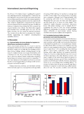
Figure 7. In vitro biological test results (solution transmittance and cell survival rate) for the smooth disc and OLS disc.
Volume 10 Issue 2 (2024) 554 doi: 10.36922/ijb.2590