
Page 65 - IJB-10-2
P. 65
International Journal of Bioprinting Advancements in 3D printing
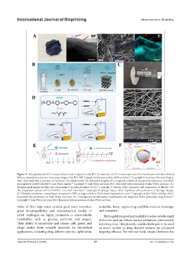
Figure 11. Polycaprolactone (PCL) and poly(lactic acid-co-glycolic acid) (PLGA) materials. (A) PCL electrospun nanofiber membranes with immobilized
69
MXene; transmission electron microscopy image of the PCL-MX-2 sample (red arrows point to MXene flakes). Copyright © American Chemical Society
2023. Reprinted with permission of American Chemical Society. (B) Schematic diagram of a composite scaffold of mesoporous hyaluronic acid (HA)
70
microspheres loaded with BMP-2 and PLGA matrix. Copyright © John Wiley and Sons 2023. Reprinted with permission of John Wiley and Sons. (C)
71
Morphological images of fabricated electrospun PCL/polycarbosilane (PCS). Copyright © Elsevier 2022. Reprinted with permission of Elsevier. (D)
72
The preparation process of PLGA/PATGP core-shell nanofibers. Copyright © Springer Nature 2022. Reprinted with permission of Springer Nature.
73
(E) Fibroblast membrane‐camouflaged nanoparticle (FNP) is supported by a PLGA-based nanoparticle cores. Copyright © John Wiley and Sons 2022.
Reprinted with permission of John Wiley and Sons. (F) Contemporary stealth surface modifications for improved PLGA particulate drug delivery.
74
Copyright © John Wiley and Sons 2022. Reprinted with permission of John Wiley and Sons.
virtue of their high water content, good water retention, materials, tissue engineering scaffolds, medical dressings,
good biocompatibility, and microstructure similar to and cosmetics.
ECM. Hydrogels are highly permeable to water-soluble Hydrogels have good permeability to water-soluble small
metabolites, such as glucose, nutrients, and oxygen. molecules and can release loaded substances continuously
Their ability to immobilize and release cells, genes, and for a long time. This property enables hydrogels to be used
drugs makes them versatile materials for biomedical as smart carriers in drug delivery systems for enhanced
applications, including drug delivery systems, ophthalmic targeting efficacy. The vitreous body, situated between the
Volume 10 Issue 2 (2024) 57 doi: 10.36922/ijb.1752