
Page 70 - IJB-10-2
P. 70
International Journal of Bioprinting Advancements in 3D printing
encapsulated in xanthan alginate gel. Liu et al. prepared To meet this demand, 3D artificial organ technology
107
PCL/SrHA composite scaffolds via 3D printing, which is harnessed to fabricate blood vessels and vascular
hold great promise as bone tissue engineering implant repair materials for transplantation. For instance, Gold
materials. Another achievement was accomplished in et al. introduced a novel nanoengineered hydrogel
108
the creation of a 3D-printed biodegradable scaffold for bioink capable of 3D-printing anatomically accurate
controlled release of deferoxamine, which is essential multicellular blood vessels by recreating the intricate
for angiogenesis and osteogenesis. The scaffold’s physical and chemical microenvironment of the human
109
design aligns with bone development and remodeling vasculature. A bioprinting strategy known as sequential
110
through surface ammonolysis and layer-by-layer printing in a reversible ink template (SPIRIT) was
assembly techniques. The combination of 3D printing developed by Fang et al., which allows the generation
technology and digital design effectively enhances internal of cardiac tissue and organoids through extensive stem
fixation and implantation outcomes for complex tibial cell proliferation and cardiac differentiation. Moore
111
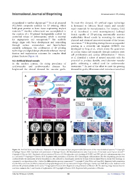
plateau fractures (Figure 14).
et al. identified a subset of natural materials with the
4.2. Artificial blood vessels potential to produce durable, small-diameter vascular
In the modern context, the rising prevalence of grafts, addressing a critical need for cardiovascular
112
cardiovascular and cerebrovascular diseases has treatments. As part of the effort to meet the growing
heightened the clinical demand for vascular grafts. demand for grafts, Silbermann et al. introduced a method
Figure 14. Artificial bone. (A) Schematic illustration for the femtosecond laser programmable fabrication of the musculoskeletal systems. Copyright
104
105
© Springer Nature 2020. Reprinted with permission of Springer Nature. (B) Simulated anterior plate fixation on a 3D-printed sacral fracture model.
Copyright © Elsevier 2021. Reprinted with permission of Elsevier. (C) Hematoxylin–eosin staining of CS-PEG and PEG gels after subcutaneous injection
in a rat model. Copyright © Elsevier 2010. Reprinted with permission of Elsevier. (D) Chondrogenic differentiation after the bioprinting process was
106
confirmed with the assessment of GAG deposition in cells cultured in chondrogenic media for 28 days (GAG is stained blue in Alcian blue staining), as
107
compared to no deposition at day 1, before the differentiation process commenced. Copyright © John Wiley and Sons 2023. Reprinted with permission
of John Wiley and Sons. (E) Microcomputed tomography images and 3D reconstruction images of Sprague-Dawley rats’ skull defects after treatment with
108
different 3D-printed scaffolds for 12 weeks. Copyright © Elsevier 2019. Reprinted with permission of Elsevier. (F) The staining image showed that the
new bone matured after 4 and 8 weeks of implantation.109 Copyright © Elsevier 2019. Reprinted with permission of Elsevier.
Volume 10 Issue 2 (2024) 62 doi: 10.36922/ijb.1752