
Page 74 - IJB-10-2
P. 74
International Journal of Bioprinting Advancements in 3D printing
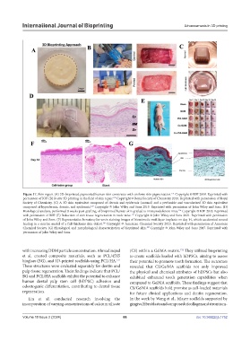
Figure 17. Skin repair. (A) 3D-bioprinted pigmented human skin constructs with uniform skin pigmentation. Copyright © IOP 2018. Reprinted with
121
permission of IOP. (B) In situ 3D printing in the field of skin repair. Copyright © Royal Society of Chemistry 2018. Reprinted with permission of Royal
122
Society of Chemistry. (C) A 3D skin equivalent composed of dermis and epidermis (normal) and a perfusable and vascularized 3D skin equivalent
123
composed of hypodermis, dermis, and epidermis. Copyright © John Wiley and Sons 2019. Reprinted with permission of John Wiley and Sons. (D)
Histological analysis, performed 8 weeks post-grafting, of bioprinted human skin grafted to immunodeficient mice. Copyright © IOP 2018. Reprinted
124
with permission of IOP. (E) Induction of skin tissue regeneration in nude mice. Copyright © John Wiley and Sons 2021. Reprinted with permission
125
of John Wiley and Sons. (F) Representative hematoxylin–eosin staining images of biomimetic multilayer implants on day 14, which accelerated wound
126
healing in a murine model of a full-thickness skin defect. Copyright © American Chemical Society 2023. Reprinted with permission of American
127
Chemical Society. (G) Histological and morphological characterization of bioprinted skin. Copyright © John Wiley and Sons 2017. Reprinted with
permission of John Wiley and Sons.
with increasing DDM particle concentration. Ahmadinejad (CS) within a GelMA matrix. They utilized bioprinting
132
et al. created composite materials, such as PCL/45S5 to create scaffolds loaded with hDPSCs, aiming to assess
bioglass (BG), and 3D-printed scaffolds using PCL/HA. their potential to promote tooth formation. The outcomes
131
These structures were evaluated separately for dentin and revealed that CS/GelMA scaffolds not only improved
pulp tissue regeneration. Their findings indicate that PCL/ the physical and chemical attributes of hDPSCs but also
BG and PCL/HA scaffolds exhibit the potential to enhance exhibited enhanced tooth generation capabilities when
human dental pulp stem cell (hDPSC) adhesion and compared to GelMA scaffolds. These findings suggest that
odontogenic differentiation, contributing to dental tissue CS/GelMA scaffolds hold promise as cell-loaded materials
regeneration. for future clinical applications and dentin regeneration.
Lin et al. conducted research involving the In the work by Wang et al., bilayer scaffolds supported by
incorporation of varying concentrations of calcium silicate gingival fibroblasts and composed of collagen and strontium-
Volume 10 Issue 2 (2024) 66 doi: 10.36922/ijb.1752