
Page 75 - IJB-10-2
P. 75
International Journal of Bioprinting Advancements in 3D printing
doped calcium silicate (SrCS) were fabricated. Achieved formation after 8 weeks. Yang et al. designed 3D-bioprinted
133
through 3D printing, these cell-supported collagen/SrCS biomimetic periodontal modules using GelMA/dECM
bilayer scaffolds demonstrated the potential to promote cell-loaded bioinks. These modules exhibited high
22
bone regeneration in the context of osteoporosis, while integrity and substantially enhanced the regeneration of
also enhancing osteogenesis. This approach is particularly periodontal tissues in Beagle hybrid models. Notably, they
relevant for periodontal regeneration. Kim et al. evaluated facilitated the development of well-aligned periodontal
the effects of biocomposites incorporated with bioactive fibers, anchoring structures at the bone-ligament interface,
cells, bone-derived dECM, and calcium phosphate and highly mineralized alveolar bone (Figure 18).
ceramics on osteogenic and odontogenic differentiation
of hDPSCs. The hDPSC-loaded biocomposite was 4.6. Cellular printing
134
implanted into the subcutaneous region of mice, leading to Cell printing pertains to incorporating living cells in
significant osteogenic/odontogenic heterotopic hard tissue biomaterials, like hydrogels, for generating bioinks. These
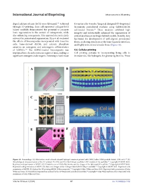
Figure 18. Stomatology. (A) Fabrication result of tooth-shaped hydrogel construct printed with DPSC-laden DDM particle bioink (10% w/v). (B)
130
Morphological characterization of the 3D-printed PCL/BG and PCL/HyA bilayer scaffolds CAD models of the scaffolds. Copyright © MDPI 2021.
131
Reprinted with permission of MDPI. (C) Printability test of CS/GelMa bioinks and the images of the fabricated 3D scaffolds. Copyright © MDPI 2021.
132
Reprinted with permission of MDPI. (D) Side-view images of the Collagen bioink/SrCS bilayer scaffolds. Copyright © MDPI 2021. Reprinted with
133
permission of MDPI. (E) Optical for 3D printing dECM-20 biocomposite. Copyright © John Wiley and Sons 2022. Reprinted with permission of John
134
Wiley and Sons. (F) Periodontal regeneration induced by the 3D-bioprinted periodontal module. Copyright © John Wiley and Sons 2023. Reprinted with
22
permission of John Wiley and Sons.
Volume 10 Issue 2 (2024) 67 doi: 10.36922/ijb.1752