
Page 71 - IJB-10-2
P. 71
International Journal of Bioprinting Advancements in 3D printing
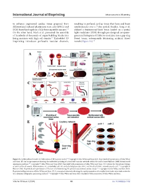
to enhance engineered cardiac tissue prepared from resulting in perfused cardiac tissue that fuses and beats
differentiated induced pluripotent stem cells (iPSCs) and synchronously over a 7-day period. Besides, Song et al.
ECM-based hydrogels in a fully biocompatible manner. utilized a femtosecond laser beam loaded on a spatial
113
On the other hand, Mark et al. pioneered the assembly light modulator (SLM) through pre-designed computer-
of hundreds of thousands of organ-building blocks into generated holograms (CGHs) to modulate into a gap-ring
living matrices with high cell density. Embedded 3D Bessel beam, subsequently fabricating artificial blood
114
bioprinting introduces perfusable vascular channels, vessels (Figure 15). 114
110
Figure 15. Artificial blood vessel. (A) Fabrication of 3D vascular model. Copyright © John Wiley and Sons 2021. Reprinted with permission of John Wiley
and Sons. (B) An image sequence showing the embedded printing of a branched vascular network within the micro-based biphasic (MB) bioink-based
111
suspension medium. Copyright © John Wiley and Sons 2023. Reprinted with permission of John Wiley and Sons. (C) Criteria for the rational design
112
of artificial blood vessels. Abbreviations: EC, endothelial cell; EG, endothelial glycocalyx; RBC, red blood cell; SMC, smooth muscle cell. Copyright ©
Elsevier 2022. Reprinted with permission of Elsevier. (D) Reinforcing 3D-printed vascularized cardiac tissue. Copyright © John Wiley and Sons 2023.
113
Reprinted with permission of John Wiley and Sons. (E) A conceptual schematic showing the rapid preparation of complex biomimetic microtube networks
114
by a dynamic holographic processing method. Copyright © John Wiley and Sons 2023. Reprinted with permission of John Wiley and Sons.
Volume 10 Issue 2 (2024) 63 doi: 10.36922/ijb.1752