
Page 67 - IJB-10-2
P. 67
International Journal of Bioprinting Advancements in 3D printing
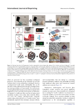
Figure 12. Hydrogel materials. (A) Photographs of sodium alginate(SA) solution, human-like collagen (H-A) solution, and formed hydrogel. Copyright
83
© Springer Nature 2023. Reprinted with permission of Springer Nature. (B) Schematic illustration and photos of the CNTs-Ecoflex0.1/PNIPAm0.5 as an
electric switch to turn on/off red and green lights under NIR irradiation. Copyright © Elsevier 2021. Reprinted with permission of Elsevier. (C) Schematic
84
diagram of the positive and negative curvatures bending for electroactive hydrogel as a function of the separated electric field with a vertical electrode
configuration. Copyright © Springer Nature 2020. Reprinted with permission of Springer Nature. (D) The structure of the electric and magnetic field
85
integrated systems. Copyright © IOP 2020. Reprinted with permission of IOP. (E) Scanning electron microscopy images of microgel@PAM/CS hydrogels
86
and typical macroscopic wound healing panorama on days 7, 11, and 15. Copyright © American Chemical Society 2023. Reprinted with permission of
87
American Chemical Society.
which are renowned for their exceptional mechanical and biocompatibility. They also emerge as a promising
properties. These materials can be fashioned into artificial interface material for artificial joints, potentially surpassing
implants, dental crowns, and patches, restoring the function both traditional alloy joints and conventional ceramic
of compromised tooth tissues. Additionally, they can be artificial joint materials (Figure 13).
utilized to create artificial joints that replace damaged joints
for restoring mobility and functionality. The continuously Furthermore, hydroxyapatite and bioactive glass
advancing biomedical ceramics preparation techniques have biomedical ceramic materials have gained significant
bequeathed ceramic artificial joints superior performance, attention in recent years. These materials find widespread
as compared to traditional alloy materials, in replacement use as coatings for metallic implants or as components in
operations of joints, such as hip, knee, shoulder, elbow, composite implants, such as bone screws, plates, and other
and wrist. For instance, alumina-zirconia composite orthopedic implants. These materials capitalize on their
ceramic joints, often referred to as “powder pottery,” have strong biocompatibility properties, leading to substantial
substantially improved wear resistance, chemical stability, improvements in the osseointegration capability of
Volume 10 Issue 2 (2024) 59 doi: 10.36922/ijb.1752