
Page 72 - IJB-10-2
P. 72
International Journal of Bioprinting Advancements in 3D printing
4.3. Artificial organ rates of bioink and crosslinker solutions, along with the
The integration of precision medicine approach into 3D speed at which a pair of rollers actively moved the cartridge
printing of human organs was first proposed by Radenkovic across the surface. Additionally, Kim et al. presented a novel
et al. This concept involves seeding cells with optimal printing platform for the creation of mature, perfusable
115
resolution, ensuring their viability during printing, vascularized 3D human skin equivalents, encompassing
123
employing compatible printers, and ultimately implanting epidermis, dermis, and subcutaneous tissue. This
the fabricated organs into patients. For instance, creating advancement holds potential for offering an enhanced in
artificial livers involves emulating hepatic lobule structure vitro platform for pathology research and the investigation
to manufacture hepatic units, the fundamental components of dermatological mechanisms. The application of 3D
of liver structure and function. Yuan et al. presented a bioprinting in regenerative medicine provides a flexible tool
robust gelatin-based hydrogel with remarkable toughness to address these challenges. For instance, Cubo utilized this
and biocompatibility, which can be directly sutured to technique to print two layers of human skin using bioinks
severed tendons of adult rabbits, rapidly promoting tendon containing human plasma, primary human fibroblasts,
124
differentiation and recovery to the initial state within 8 and keratinocytes obtained from skin biopsies. This
weeks. In another study, Wang et al. developed a clinical- highlights the potential of 3D bioprinting in generating
116
grade bioartificial liver (BAL) device utilizing human skin constructs for various applications, ranging from
induced hepatocytes (hiHeps) produced under Good regenerative medicine to dermatological research.
Manufacturing Practice (GMP) conditions. Meanwhile, Ma et al. employed “cell writing” bioprinting
117
Liu et al. successfully repaired a skull model with a sagittal technology to create a functional skin substitute, supplied
suture defect. Several studies have proposed techniques with blood vessels, based on a biomimetic multicellular
118
based on 3D printing for facilitating artificial organ system. This system, encompassing SS-containing
125
creation. John et al. reported a method for creating aligned constructs, showcased an unprecedented blend of
cardiac tissue using anisotropic organ building blocks vascularized skin-like structure and the capacity to induce
bioprinted from human-induced pluripotent stem cell- vascularization. It demonstrated remarkable angiogenic
derived cardiomyocytes. This approach enables high cell activity both in vitro and in vivo. This investigation offers
119
density and intricate cell arrangements, which are critical insights into crafting biomimetic multicellular structures
determinants for functional cardiac tissue creation. On the with angiogenesis-inducing attributes, which can aid in
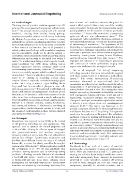
other hand, Noor et al. presented a simple technique for regenerating vascularized complex and layered tissues.
3D printing thick, vascularized, perfusable cardiac patches Zhang et al. developed a swift and straightforward method
tailored to a patient’s immune, cellular, biochemical, to directly process adipose tissue into microfragmented
and anatomical attributes. Mathematical modeling of adipocyte ECM. This matrix was harnessed as the
120
126
oxygen delivery further improved vascular structure, and primary component of bioinks for fabricating biomimetic
the elongated cardiomyocytes in these patches exhibited multilayered implants via 3D bioprinting. This approach
abundant actin striations (Figure 16). accelerates the production of bioprinted skin substitutes
and holds promise for treating full-thickness skin defects.
4.4. Skin repair Puchett et al. showcased newly formulated inks and the
Researchers have explored various ECMs in the context capability to generate comprehensive skin models through
of 3D printing technology for skin applications. This an open-source printer. Through immunostaining and
127
approach aims to optimize the natural properties and electron microscopy, they demonstrated that the bioprinted
activity of skin, allowing for effective repair of damaged skin displayed all the characteristics of human skin at both
skin post-transplantation and seamless integration molecular and macromolecular levels. This underscores
between printed and normal skin. Wu et al. utilized three the potential of bioprinting to create highly authentic skin
distinct skin cell types—keratinocytes, melanocytes, and models for various applications (Figure 17).
fibroblasts from different donors—to create a 3D-colored
human skin structure that closely resembles the complex 4.5. Stomatology
pigmentation of the donors. This innovation has enabled Mouthwash tools are made of a variety of materials,
121
the development of in vitro 3D skin constructs with including alloys, biochemical ceramics, and photosensitive
coloration, serving purposes such as toxicology testing resins. Alloys are predominantly used in 3D printing for
and basic cell biology research. Hakimi et al. introduced creating toothed belts, dental bridges, metal inner crowns,
a handheld skin printer capable of on-site fabrication and other components. Laser sintering is sometimes
of biomaterial and skin tissue sheets featuring varied employed in tooth restoration. Ceramic 3D printing
homogeneity and structural composition. Precise sheet is used to repair damaged oral tools and restore gear
122
formation was achieved by synchronizing the delivery flow components, especially for backlash correction, and is
Volume 10 Issue 2 (2024) 64 doi: 10.36922/ijb.1752