
Page 54 - IJB-3-2
P. 54
Hybrid three-dimensional (3D) bioprinting of retina equivalent for ocular research
Figure 2. Cell viability assay of manually seeded and bioprinted ARPE-19 cells, p > 0.05
Figure 3. Fluorescent images (F-actin) of bioprinted ARPE-19 cells at day 1 (a), day 7 (b) and day 14 (c); scale bar: 200 µm
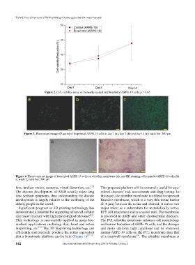
Figure 4. Phase-contrast image of bioprinted ARPE-19 cells on ultrathin membrane (a), and HE staining of bioprinted ARPE-19 cells (b)
at week 2; scale bar: 500 µm
loss, unclear vision, scotoma, visual distortion, etc. [20] This proposed platform will be extremely useful for eye-
The disease development of AMD usually takes long related diseases’ risk assessments and drug testing. In
time without symptoms, thus understanding the disease this paper, the ultrathin membrane is utilized to represent
development is largely relative to the wellbeing of the Brunch’s membrane, which is a very thin tissue barrier
elderly people in the world. (2–4 µm) between the retina and choroid. It serves two
Significant progress in 3D printing technology has major roles: as a substratum for metabolically active
demonstrated a potential for organizing advanced cellular RPE cell attachment and as a vessel wall. The membrane
and tissue structure with high physiological relevance . is involved in AMD and other chorioretinal diseases.
[21]
This technology is successfully applied in many bio- The PCL ultrathin membrane enhances cell morphology
medical applications including skin, heart and retina and barrier formation of ARPE-19 cells, and the stronger
bioprinting, etc. [22] The 3D bioprinting technology can and more uniform tight junctions can be observed
efficiently and precisely produce the retina equi valent among ARPE-19 cells on the PCL membrane than that
that a biomimetic platform can be built (Figure 1) [23–25] . of a transwell membrane . The ultrathin membrane is
[16]
142 International Journal of Bioprinting (2017)–Volume 3, Issue 2