
Page 56 - IJB-3-2
P. 56
Hybrid three-dimensional (3D) bioprinting of retina equivalent for ocular research
Figure 8. Live/dead assay of Y79 cell in bioprinted alginate/pluronic complex bioink at day 1 (a), day 4 (b) and day 7 (c); scale bar: 200
µm
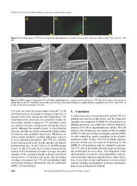
Figure 9. SEM images of bioprinted Y79 cell-laden alginate/pluronic complex bioink (a, b and c); Y79 cell distribution at the surface of
bioink (d), and the Y79 cell distribution at the cross sections of the alginate/pluronic complex bioink at magnification of 200× and 1000× (e
and f); yellow arrows indicate Y79 cells
[35]
Y79 cells are useful for retina-related research in 3D 5. Conclusion
cell-bioprinting to investigate biological responses of
sensory retina cells during and after bioprinting. The A retina equivalent was prepared by hybrid 3D bio-
bioprinted retina constructs are cultured in media for printing and reported in this article, and the bioprinted
seven days, and the bioprinted Y79 cell-laden bioink construct was composed of ARPE-19 cell monolayer on
can preserve its basic configuration during the culture ultrathin membrane, and a third layer with two distinctive
period. Although the complex bioink is crosslinked by patterns (Y79 cells in alginate/pluronic bioink). The cell
calcium ions that are slowly released in culture media, viability, cell morphology and quality of the bioprinted
its structure may gradually deteriorate. Therefore, the ARPE-19 cells were closely investigated, and the ARPE-
culture media should be carefully and gently removed 19 cells formed high quality monolayer on the ultrathin
to avoid structural disruption. The Y79 cell viability membrane within 14 days. The Y79 cell-laden alginate/
is not compromised in the bioink, and the cell density pluronic bioink was successfully bioprinted on the
increases from day 1 to day 7 (Figure 8). In SEM images ARPE-19 cell monolayer with two distinctive patterns.
(Figure 9), the Y79 cells can be easily observed on the The Y79 cells in the bioink showed benign morphology
surface of the bioprinted cell-laden hydrogel; the inner and proliferated in seven days. The bioprinted retina
structure of the cell-laden bioink is porous and allows equivalent has acceptable cytocompatibility with ad-
transportation of nutrients and waste, thus providing vanced structure aiming to simulate native retina. There-
a benign environment for Y79 cell reproduction while fore, it may have broad applications in retina-related
maintaining biological interactions with the underneath research including investigations on disease mechanism,
ARPE-19 cell monolayer. drug testing and treatment methods.
144 International Journal of Bioprinting (2017)–Volume 3, Issue 2