
Page 156 - IJB-10-3
P. 156
International Journal of Bioprinting 3D bone: Current & future
become impervious to nutrients, and the chondrocytes 2.3.2. Stem cells and bone differentiation
inside the cartilage begin to die, thereby allowing blood Stem cells are undifferentiated cells that develop into
vessels to enter the empty cavity (primary ossification specific cells of other tissues through specific differentiation
center) through the periosteum. Osteoprogenitor pathways. These cells can be either embryonic, adult,
20
22
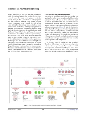
cells are transported through blood vessels from the or induced pluripotent stem cells, and based on their
primary ossification center toward the ends of the developmental potential, they can be divided into five
bones and replace the cartilage tissues with bone tissues groups: totipotent, pluripotent, multipotent, oligopotent,
(endochondral ossification), forming a spongy bone. 19,20 and unipotent stem cells (Figure 5). After fertilization, the
After the cartilage model is completely replaced in the zygote of a single cell begins to divide (barring). Totipotent
diaphysis, the osteoclasts open the medullary cavity inside stem cells refer to actively dividing undifferentiated stem
the bone. Meanwhile, in the epiphyses, chondrocytes cells (i.e., less than 16 cells [morula]) and are capable of
19
proliferate and the cartilages continue to grow, forming a forming cells of any tissue. The morula then develops into
secondary ossification center. At the end of the process, the a blastocyte, where the cells are pluripotent. Consequently,
entire cartilage model is replaced by bone tissues except these cells can no longer form extraembryonic tissue but
for the epiphyseal plate, located between the epiphysis and can form germinal cells and gametes.
the diaphysis, and this facilitates subsequent longitudinal
bone growth by endochondral ossification. While the In the next stage of development, the monolayer
interior of the cartilage model is replaced by spongy bone, blastoderm differentiates into the three germ layers: the
the perichondrium transforms into the periosteum and endoderm (inner germ layer), the mesoderm (middle
the surface osteoblasts form the compact bone. These two germ layer), and the ectoderm (outer germ layer). These
types of bone growth continue until about 20–25 years, cells are also pluripotent and form the cells of the different
after which only bone regeneration occurs. tissues. The endoderm comprises epithelial cells from the
Figure 5. Types of embryonic stem cells. This figure was created using BioRender.
Volume 10 Issue 3 (2024) 148 doi: 10.36922/ijb.2056