
Page 274 - IJB-10-3
P. 274
International Journal of Bioprinting 3D bioscaffolds with SR1 for vasculogenesis
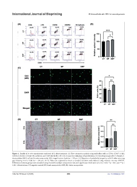
Figure 1. Results of in vitro experiments conducted 48 h after treatment. (A) Flow cytometry analysis using antibodies such as CD34, CD117 (c-kit),
CD184 (CXCR4), CD144 (VE-cadherin), and VEFGR2 (KDR). (B) CCK-8 assay for evaluating cell proliferation. (C) Resulting images after 5-ethynyl-2’-
deoxyuridine (EdU) cell proliferation assay under 200× magnification. Scale bar = 100 µm. (D) Migration of endothelial progenitor cells 8 h after removing
gap-inducing inserts. Scale bar = 250 µm. (A–D) Values are expressed as mean ± standard deviation and analyzed using ordinary one-way ANOVA.
Differences between groups were analyzed using Dunnett’s multiple comparisons test, and significance levels were set at *p < 0.05, **p < 0.01, and ***p <
0.001. Abbreviations: CT, negative control; NP, blank nanoparticle; SNP; SR1-laden nanoparticle.
Volume 10 Issue 3 (2024) 266 doi: 10.36922/ijb.1931