
Page 278 - IJB-10-3
P. 278
International Journal of Bioprinting 3D bioscaffolds with SR1 for vasculogenesis
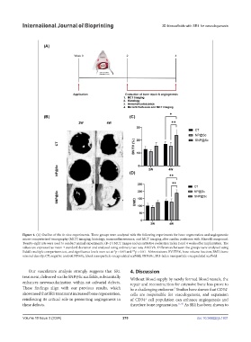
Figure 4. (A) Outline of the in vivo experiments. Three groups were analyzed with the following experiments for bone regeneration and angiogenesis:
micro-computerized tomography (MCT) imaging, histology, immunofluorescence, and MCT imaging after cardiac perfusion with Microfil compound.
Twenty-eight rats were used to conduct animal experiments. (B–D) MCT images and quantitative evaluation index 2 and 4 weeks after implantation. The
values are expressed as mean ± standard deviation and analyzed using ordinary two-way ANOVA. Differences between the groups were analyzed using
Sidak’s multiple comparisons test, and significance levels were set at *p < 0.05 and **p < 0.01. Abbreviations: BV/TV%, bone volume fraction; BMD, bone
mineral density; CT, negative control; NP@Sc, blank nanoparticle-encapsulated scaffold; SNP@Sc, SR1-laden nanoparticle-encapsulated scaffold.
Our vasculature analysis strongly suggests that SR1 4. Discussion
treatment, delivered via the SNP@Sc scaffolds, substantially Without blood supply by newly formed blood vessels, the
enhances neovascularization within rat calvarial defects. repair and reconstruction for extensive bone loss prove to
These findings align with our previous results, which be a challenging endeavor. Studies have shown that CD34
+
7
showcased that SR1 treatment increased bone regeneration, cells are responsible for vasculogenesis, and expansion
reinforcing its critical role in promoting angiogenesis in of CD34 cell population can enhance angiogenesis and
+
these defects. therefore bone regeneration. 11,35 As SR1 has been shown to
Volume 10 Issue 3 (2024) 270 doi: 10.36922/ijb.1931