
Page 292 - IJB-10-3
P. 292
International Journal of Bioprinting Design and optimization of 3DP bioscaffolds
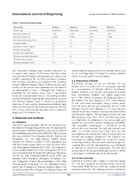
Table 2. Grid partitioning settings
Grid setting Domain 1 Domain 2 Domain 3 Domain 4
Element type Hexahedral Tetrahedral Hexahedral Hexahedral
Maximum element size 1.000 0.189 0.800 1.000
Minimum element size 0.1800 0.0204 0.100 0.1800
Maximum element growth rate 1.5 1.1 1.45 1.5
Curvature factors 0.6 0.4 0.5 0.6
Resolution of narrow regions 0.5 0.9 0.6 0.5
Number of sweep units 30 - 40 30
Number of boundary layers 8 8 5 8
Boundary layer stretching factors 1.2 1.2 1.1 1.2
Thickness of the first layer 1 1 1 1
Thickness adjustment factors 0.10 0.10 0.08 0.10
and convection–diffusion mass transfer dominate, can culture dishes (Corning Life Sciences, Suzhou, China), and
be sparser and coarser. Furthermore, boundary layers 15 mL centrifuge tubes (Corning Life Sciences, Suzhou,
were introduced during mesh generation to enhance the China) were also used in the experiments.
model’s convergence for the fluid calculation processes,
and transition smoothing was required at the interfaces 3.2. Preparation of bioink
between different domains. Figure 3b showcases the mesh The bioink consisted of 5% w/v PEGDA, 5% w/v
results, and the detailed mesh parameters for each domain GelMA, 0.5% w/v LAP, 0.03% w/v tartrazine, and cells
are summarized in Table 2. Although finer meshing is at a concentration of 300,000 cells/mL. Specifically,
preferable for the porous media flow, it significantly GelMA, PEGDA, LAP powder, and tartrazine powder
lowers the computational efficiency. The mesh generation were individually weighed and added sequentially
quality was tuned by adjusting the computational accuracy into a PBS solution to prepare the hydrogel solution.
and efficiency balance. Figure 3c shows the quantitative The solution was then heated in a 37°C water bath for
depiction of mesh quality, demonstrating relatively high 15 min and mixed thoroughly using a vortex mixer.
values. The time step for the model was set at interval of (0, Once the above process was completed, the pH of the
0.01, 7) days, where 0 is the start time, 0.01 is a time step hydrogel solution was adjusted to 7.4 using standard
length, and 7 is the finial time. sodium bicarbonate titration solution and hydrochloric
acid titration solution. The hydrogel solution was then
3. Materials and methods filtered using a filter. The C2C12 cells that had grown
to a high level of confluence in the culture dish were
3.1. Materials added. First, the culture medium (high-glucose DMEM:
The C2C12 mouse myoblast cell line was obtained from FBS: penicillin: streptomycin = 100:10:1:1) in the
the School of Biology, Hunan University. The materials for culture dish was removed. The dish was washed twice
generating the scaffold were gelatin methacryloyl (GelMA) with 2 mL of PBS solution for 5 min. Then, the cells
and polyethylene glycol diacrylate (PEGDA). The tartrazine were digested with trypsin for 4 min to ensure both cell
dye was purchased from Shanghai Aladdin Biochemical viability and complete detachment. The digestion was
Technology Co., Ltd. High glucose DMEM culture terminated by adding double the volume of trypsin-
medium, 1% antibiotic solution, 2 mM L-glutamine, and containing medium. The cells were counted using a cell
10% fetal bovine serum (FBS) were obtained from Gibco counting plate, and the cell suspension was centrifuged
Life Technologies (Waltham, MA, USA). 4’,6-Diamidino- at 1000 rpm to remove the supernatant. The cells were
2-phenylindole (DAPI) and Cell Counting Kit-8 (CCK-8) resuspended in the prepared hydrogel solution to obtain
were purchased from Beyotime Biotechnology (Shanghai, a cell density of 300,000 cells/mL for the bioink.
China) Co., Ltd. Phosphate-buffered saline (PBS) was
obtained from Thompson Biological Technology Co., 3.3. In vitro culturing
Ltd. (Hangzhou, China). The photoinitiator LAP (Bide Cylindrical scaffolds with a radius of 2 mm and height
Pharmaceutical Technology Co., Ltd., Shanghai, China), of 2 mm were designed and printed using a nanoArch
pipette tips (Corning Life Sciences, Suzhou, China), 0.22 S140 printer (BMF Precision Tech Inc.) using the bioink
μm sterile syringe filters (Dragon Lab, Beijing, China), cell prepared previously. The printing process is illustrated
Volume 10 Issue 3 (2024) 284 doi: 10.36922/ijb.1838