
Page 295 - IJB-10-3
P. 295
International Journal of Bioprinting Design and optimization of 3DP bioscaffolds
single channel in the scaffold. Specifically, as shown in the 4.1. Model validation and modeled physical fields
flowchart (see Figure 5), the initial volume fraction of the To examine the accuracy of the multi-physics model in
central channel in the scaffold was set to 2.5%. Iterative predicting cell growth, a series of experiments involving
calculations were performed using the multi-physics model C2C12 cell cultivation, bioink preparation, DLP
with an increment of volume fraction. The optimal volume manufacturing of cell-laden scaffolds, dynamic cultivation
fraction of the channel was then obtained when the average of scaffolds, and cell proliferation assays were conducted.
cell density and the total amount of cells in the scaffold no The C2C12 mouse myoblast cell line was selected because
longer increased. In the second step, the single-channel it is often used in pharmaceutical science and biomedical
scaffold was transformed into a uniformly distributed multi- research to examine tissue metabolism and cell growth,
channel structure with equivalent void volume fraction, and is widely used for validating cell growth models. 22,29 In
where the optimal channel diameter and number values addition, the cell kinetic parameters of C2C12 required to
were obtained by iterative calculations. drive the model have been well-established in many studies
(see Table 4). The comparison between the simulated and
4. Results and discussion experimental cell growth trends in cylindrical scaffolds
The modeling system was validated by comparing the without channels within 7 days is depicted in Figure 6,
modeled data to those observed to demonstrate its exhibiting reasonably good agreement. Cell proliferation
ability to predict cell growth and then applied to analyze was increased by 4 times on day 7, as shown by both model
the spatiotemporal behaviors of the associated physical and experimental results. The maximum discrepancy
fields. The parametric study was subsequently performed between the model and experiments was 4.38%, which
to gain insight into the mechanism of how geometric occurred on day 4, likely due to the sensitivity issues of the
and culturing parameters influence cell growth. Finally, detection reagents or the simplified model physics, such
a showcase of optimizing the design of channeled as the exclusion of biological forces and cell death factors.
scaffolds was presented to demonstrate the effectiveness The selection of the model parameters can also contribute
of the two-step optimization method proposed in to the observed discrepancy (see section 4.2). Based on its
this study. well-modeled cell growth trend and relatively small error,
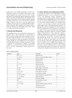
Table 4. Model parameters
Parameters Values Description Sources
Q 600 μL ∙ min -1 Initial flow rate Set
0
-4
μ 8.4 × 10 Pa∙s Dynamic viscosity of culture medium 36
ρ 1009 kg∙m -3 Nutrient density 36
ε 0 0.7 Initial porosity 20
Γ 3.45 Initial tortuosity 30
0
-12
κ 0 6.76 ×10 m 2 Initial permeability 30
P 0Pa Outlet static pressure Set
0
C 0 222.5 μM Initial oxygen concentration 20
C 222.5 μM Inlet oxygen concentration 20
in
-9
D 0 3.10 × 10 m ∙s -1 Initial diffusion coefficient 30
2
K 0.006 mol∙m -3 M-M equation constant 37
m
V 3.0 × 10 mol∙cell ∙s -1 Actual maximum oxygen uptake rate 20
-17
-1
r
ρ 3.0 × 10 cells∙cm 3 Initial cell density Set
-6
0
ρ 1020 kg∙m -3 Cell volume density 30
m
K 0.006 Contois equation constant 38
c
V 7.24 × 10 m 3 Cell volume 39
-15
c
-5
μ r 1.53 × 10 cells∙s -1 Actual maximum specific growth rate 30
t 3 h Cell adhesion period 40
0
t 1 72 h Cell spreading period 41
Volume 10 Issue 3 (2024) 287 doi: 10.36922/ijb.1838