
Page 411 - IJB-10-3
P. 411
International Journal of Bioprinting In situ thermal monitoring in bioprinting
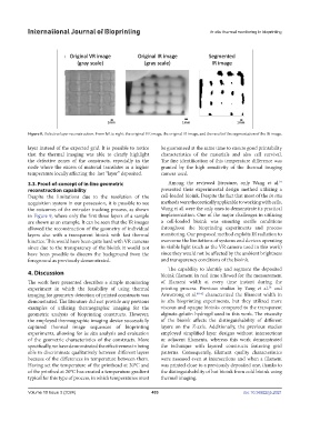
Figure 8. Defective layer reconstruction. From left to right: the original VR image, the original IR image, and the result of the segmentation of the IR image.
layer instead of the expected grid. It is possible to notice be guaranteed at the same time to ensure good printability
that the thermal imaging was able to clearly highlight characteristics of the materials and also cell survival.
the defective zones of the constructs, especially in the The fine identification of this temperature difference was
node where the excess of material translates in a higher granted by the high sensitivity of the thermal imaging
temperature locally affecting the last “layer” deposited. camera used.
34
3.3. Proof-of-concept of in-line geometric Among the reviewed literature, only Wang et al.
reconstruction capability presented their experimental design method utilizing a
Despite the limitations due to the resolution of the cell-loaded bioink. Despite the fact that most of the in situ
acquisition system in our possession, it is possible to see methods were theoretically applicable to working with cells,
the outcomes of the extruder tracking process, as shown Wang et al. were the only ones to demonstrate its practical
in Figure 9, where only the first three layers of a sample implementation. One of the major challenges in utilizing
are shown as an example. It can be seen that the IR images a cell-loaded bioink was ensuring sterile conditions
allowed the reconstruction of the geometry of individual throughout the bioprinting experiments and process
layers also with a transparent bioink with fast thermal monitoring. Our proposed method exploits IR radiation to
kinetics. This would have been quite hard with VR cameras overcome the limitations of systems and devices operating
since due to the transparency of the bioink it would not in visible light (such as the VR camera used in this work)
have been possible to discern the background from the since they would not be affected by the ambient brightness
foreground as previously demonstrated. and transparency conditions of the bioink.
The capability to identify and segment the deposited
4. Discussion bioink filament in real time allowed for the measurement
The work here presented describes a simple monitoring of filament width at every time instant during the
34
experiment in which the feasibility of using thermal printing process. Previous studies by Yang et al. and
imaging for geometry detection of printed constructs was Armstrong et al. 40-42 characterized the filament width in
demonstrated. The literature did not provide any previous in situ bioprinting experiments, but they utilized more
examples of utilizing thermographic imaging for the viscous and opaque bioinks compared to the transparent
geometric analysis of bioprinting constructs. However, alginate-gelatin hydrogel used in this work. The viscosity
the employed thermographic imaging device successfully of the bioink affects the distinguishability of different
captured thermal image sequences of bioprinting layers on the Z-axis. Additionally, the previous studies
experiments, allowing for in situ analysis and evaluation employed simplified layer designs without intersections
of the geometric characteristics of the constructs. More or adjacent filaments, whereas this work demonstrated
specifically, we have demonstrated the effectiveness in being the technique with layered constructs featuring grid
able to discriminate qualitatively between different layers patterns. Consequently, filament quality characteristics
because of the differences in temperature between them. were assessed even at intersections and when a filament
Having set the temperature of the printhead at 30°C and was printed close to a previously deposited one, thanks to
of the printbed at 20°C has created a temperature gradient the distinguishability of hot bioink from cold bioink using
typical for this type of process, in which temperatures must thermal imaging.
Volume 10 Issue 3 (2024) 403 doi: 10.36922/ijb.2021