
Page 427 - IJB-10-3
P. 427
International Journal of Bioprinting Expanding 3D cell proliferation with DLP bioprinting
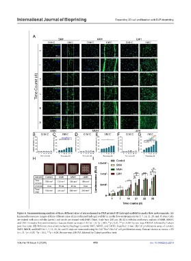
Figure 6. Immunostaining analysis of three different sizes of microchannel in DLP-printed 3D hydrogel scaffold in media flow environments. (A)
Immunofluorescence images of three different sizes of microchannel hydrogel scaffold in media flow environments for 5, 7, 14, 21, 28, and 35 days. Cells
are stained with an α-tubulin (green), and nuclei are stained with DAPI (blue). Scale bars: 200 µm. (B–G) α-tubulin confluency analysis of SMH, MMH,
and LMH in media flow environments. Data are shown as means ± SD (n = 3). *p < 0.05, **p < 0.01, ***p < 0.001 by one-way ANOVA followed by Tukey’s
post-hoc tests. (H) Different sizes of microchannel hydrogel (control, SMH, MMH, and LMH). Scale bar: 2 mm. (I) Cell proliferation assay of control,
SMH, MMH, and LMH for 1, 7, 14, 21, 28, and 35 days are measured using the Cell Titer -Glo 3.0 cell proliferation assay. Data are shown as means ± SD
®
®
(n = 3). *p < 0.05, **p < 0.01, ***p < 0.001 by one-way ANOVA followed by Tukey’s post-hoc tests.
Volume 10 Issue 3 (2024) 419 doi: 10.36922/ijb.2219