
Page 428 - IJB-10-3
P. 428
International Journal of Bioprinting Expanding 3D cell proliferation with DLP bioprinting
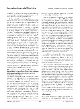
behavior of the cells at 60 rpm and 90 rpm for 14 days, we statistically significant difference between day 35 and day
observed a significant increase in cell proliferation with the 1 was observed (p < 0.001; Figure 7C).
rising rpm (Figure S2 in Supplementary File).
Based on DAPI analysis, the number of cells increased
To observe the differences in cell proliferation over each during the entire time course to 286 ± 25.71, 290 ± 58.13,
time course of the three sizes of microchannel hydrogels 366 ± 38.55, 376 ± 35.73, 401 ± 50.57, and 464 ± 60.10, with
and the effect of the microchannels, we performed cell a statistically significant difference between day 1 and day
luminescence-based quantification. The volume was 35 (p < 0.01; Figure 7D). The side view of the microchannel
fixed at 128 mm to equalize the number of cells in each cross-section shows that the cells adhered to a circular
3
hydrogel (Figure 6H). Statistical analysis of the differences shape and formed a network (Figure 7E). The analysis was
between each time course showed no significant difference performed based on the ratio of the remaining area to the
in all groups compared to the control group on day 1 initial microchannel space, showing 91.31 ± 0.06% of the
(ns). There was a statistically significant difference in the inner space area of the channel on day 1 and 64.10 ± 0.06%
luminescence levels between the MMH and LMH groups on day 7, a sharp decrease with statistical significance (p <
compared to the control group on day 7 (*b < 0.05). A 0.001). Then, the remaining space area decreased by around
significant increase was observed in the LMH group 40%, showing 64.10 ± 0.06% and 60.21 ± 0.04% of the space
compared to the control group on day 14 (*c < 0.05). All area on day 14 and day 21, respectively, with no significant
three groups showed a significant difference from the difference between them (ns). As time progressed to day
control group on day 21 (*d < 0.05, **d < 0.01, and *ddd < 21 and day 28, cells filled the unoccupied space inside the
0.001). Additionally, all three groups showed a significant channel, and the area of the remaining space decreased
increase on day 28 compared to the control group (***e sharply again from 59.07 ± 0.03% to 33.76 ± 0.10% (p <
< 0.001). Throughout the time course, LMH exhibited 0.01). Finally, on day 35, 28.80 ± 0.05% of the inner space
the highest luminescence levels. Despite having a small area of the channel was filled, leaving approximately 30%
surface area, LMH showed the highest luminescence level, of the printed microchannel shape unoccupied due to cell
indicating high cell viability and proliferation due to the proliferation from long-term culture (Figure 7F).
presence of media supply within the channel (Figure 6I). It is well known that a high-flow culture environment
Our results suggest that microchannels not only increase is more effective than a non-shaking culture in promoting
surface area but also function as a conduit to supply oxygen cell proliferation and maturation in vitro. In addition, it
27
and nutrients to encapsulated cells and facilitate media has been reported that aligned microchannels in printed
flow. Thus, the introduction of microchannels into DLP- 3D hydrogel scaffolds affect the spatial migration and
printed 3D hydrogel scaffolds, along with the introduction survival of neural stem cell spheroids. 69
of media flow, enhances cell proliferation compared to Here, we observed that cell proliferation resulted in the
the same volume of scaffold cultured under non-shaking formation of a single layer of cells in the microchannel space
culture conditions during long-term culture.
during the early-time cultivation (until day 7), whereas
3.7. Analysis of cell morphology and proliferation two or more layers of cells were formed during the mid-
enhancement by geographic location of DLP-printed and late-time cultivation (until day 21 and day 35; Videos
3D hydrogel scaffolds with multichannel (LMH) S1–S3 in Supplementary File). However, contrary to our
during long-term culture expectation that cells would completely fill the empty space
The purpose of this study was to introduce LMH, which inside the microchannel, we found that the flow of media
has been proven to be effective in cell proliferation, and provided a continuous supply of nutrients and oxygen to
to observe and analyze cell morphology, viability, and the cells without blocking the channel. This ensured the
proliferation following the geographic location (top effectiveness of introducing vertical multichannel arrays
and side views) of scaffolds (Figure 7A). We conducted into the DLP-printed 3D hydrogel scaffolds and changing
immunocytochemistry staining using α-tubulin/DAPI the culture environment to enhance cell proliferation.
and observed the top view of the cells to assess their Ultimately, this method provides a fast, simple, and efficient
morphology and proliferation. Over time, the cells means of significantly improving cell seeding, distribution,
elongated and proliferated, forming multiple layers, and proliferation in various geographical aspects (top and
which led to an increase in α-tubulin/DAPI (Figure 7B). side) during long-term culture.
The percentage of cells exhibiting α-tubulin confluency
consistently increased throughout the experiment, with 4. Conclusion
values of 14.89 ± 2.45%, 22.28 ± 3.14%, 24.42 ± 2.83%, In this study, we aimed to enhance the survival and
32.35 ± 2.53%, 37.29 ± 2.54%, and 43.46 ± 10.46%. A proliferation of cells encapsulated within DLP-printed
Volume 10 Issue 3 (2024) 420 doi: 10.36922/ijb.2219