
Page 27 - IJB-4-1
P. 27
Fan Liu, et al.
differentiation. As a follow-up study, Liu et al. investigated the
cellular niche by tailoring the architecture of a tissue construct
via cell bioprinting [139] . The change of the geometry and
architecture, such as the pore size of the tissue construct, has
a strong influence on guiding sweat-gland morphogenesis
and function [140] . The studies demonstrate that it is possible
to print a bioartificial skin with the sweat-gland regenerative
capability.
4. Conclusion
The advent of 3D bioprinting technologies has led to a
significant progress in the manufacture of large bioartificial
organs, such as the bones, livers, hearts, cartilages
and skins, with heterogenic compositions. Various
bioprinting techniques have provided a fully automated
and advanced platform to deposit multiple cell types and
ECM-like biomaterials to simulate the natural organs, a
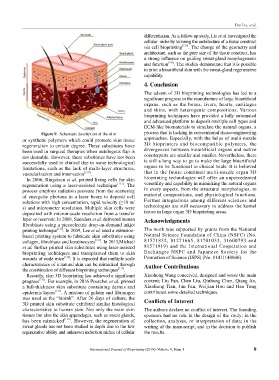
Figure 9. Schematic description of the skin process that is lacking in conventional tissue-engineering
or synthetic polymers which could promote skin tissue approaches. Especially, with the helps of multi-nozzle
regeneration to certain degree. These substitutes have 3D bioprinters and biocompatible polymers, the
been used in surgical therapies when autologous flap is divergences between bioartificial organs and native
not desirable. However, these substitutes have not been counterparts are smaller and smaller. Nevertheless, there
successfully used in clinical due to some technological is still a long way to go to make the large bioartificial
limitations, such as the lack of multi-layer structures, organs to be functional in clinical trials. It is believed
vascularization and innervation [130] . that in the future combined multi-nozzle organ 3D
In 2006, Ringeisen et al. printed living cells for skin bioprinting technologies will offer an unprecedented
regeneration using a laser-assisted technique [131] . The versatility and capability in mimicking the natural organs
process employs radiation pressure from the scattering in every aspects, from the structural morphologies, to
of energetic photons in a laser beam to deposit cell material compositions, and physiological functions.
solutions with high concentration, rapid velocity (≥10 m/ Further integrations among different sciences and
s) and micrometer resolution. Multiple skin cells were technologies are still necessary to address the kernel
deposited with micron-scale resolution from a transfer issues in large organ 3D bioprinting areas.
layer or reservoir. In 2008, Saunders et al. delivered human Acknowledgments
fibroblasts using a piezoelectric drop-on-demand inkjet
printing technique [132] . In 2009, Lee et al. used a extrusion- The work was supported by grants from the National
based printing system to fabricate skin substitutes using Natural Science Foundation of China (NSFC) (No.
collagen, fibroblasts and keratinocytes [133] . In 2013,Michael 81571832, 81271665, 81701033, 31600793 and
et al. further printed skin substitutes using laser-assisted 81571919) and the International Cooperation and
bioprinting techniques and transplanted them to skin Exchanges NSFC and Japanese Society for the
wounds of nude mice [134] . It is expected that multiple scale Promotion of Science (JSPS) (No. 81411140040).
characteristics of a natural skin can be mimicked through Author Contributions
the combination of different bioprinting techniques [135] .
Recently, skin 3D bioprinting has achieved a significant Xiaohong Wang conceived, designed and wrote the main
progress [136] . For example, in 2016 Pourchet et al. printed content; Liu Fan, Chen Liu, Qiuhong Chen, Qiang Ao,
a full-thickness skin substitute containing dermis and Xiaohong Tian, Jun Fan, Weijian Hou and Hao Tong
epidermis layers [137] . A mixture of gelatin and fibrinogen contributed some detailed techniques.
was used as the “bioink”. After 26 days of culture, the Conflicts of Interest
3D printed skin substitute exhibited similar histological
characteristics to human skin. Not only the main skin The authors declare no conflict of interest. The founding
tissues but also the skin appendages, such as sweat glands, sponsors had no role in the design of the study; in the
has been mimicked [138] . However, the regeneration of collection, analyses, or interpretation of data; in the
sweat glands has not been studied in depth due to the low writing of the manuscript, and in the decision to publish
regenerative ability and unknown induction niches of cellular the results.
International Journal of Bioprinting (2018)–Volume 4, Issue 1 9