
Page 25 - IJB-4-1
P. 25
Fan Liu, et al.
For liver 3D bioprinting, two essential elements should been developed to improve the functionality of the cardiac
be addressed: (1) a extrusion-based multi-nozzle 3D tissues. For example, in 2007, Marga et al. emitted a stream
bioprinter with an appropriate soft/hardware. (2) multiple of cell-laden hydrogel microparticles in a well-defined
cell lineages from the liver or stem cells with proper topological pattern to form 3D myocardial patches using
growth factors. The extrusion-based multi-nozzle 3D a inkjet-based bioprinting technique [110] . This technique
bioprinter can print multiple cell types along with other is supported by the self-assembly and self-organizing
biomaterials simultaneously in a layer-by-layer manner, capabilities of cells. In 2011, Gaebel et al. patterned human
which offers a great opportunity in manufacturing the stem cells and endothelial cells with laser printing for
complicated bioartificial livers with more than 6 cell cardiac regeneration [111] . In 2012, human cardiomyocyte
types or tissues [102–106] . These technologies allow to use progenitor cells (HCMPCs) in alginate hydrogel was
multiple polymeric hydrogels and growth factors to printed by the same group [112] . HCMPCs in the alginate
control the spatial distribution of cells and bioactive hydrogel showed an increase of cardiac commitment while
agents. at the same time maintaining viability and proliferation. In
3.3 Heart 3D Bioprinting 2013, Duan et al. constructed trileaflet valve like conduits
using sinus smooth muscle cells (SMCs) and alginate/
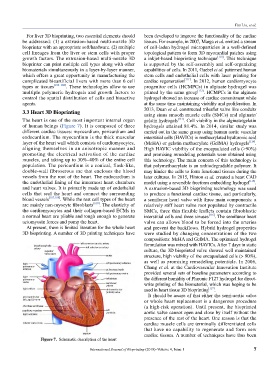
The heart is one of the most important internal organ gelatin hydrogels [113] . Cell viability in the alginate/gelatin
of human beings (Figure 7). It is composed of three hydrogels attained 81.4%. In 2014, similar study was
different cardiac tissues: myocardium, pericardium and carried out in the same group using human aortic vascular
endocardium. The myocardium is the thick muscular interstitial cells (HAVICs) in methacrylated hyaluronic acid
layer of the heart wall which consists of cardiomyocytes, (MeHA) or gelatin methacrylate (GelMA) hydrogels [114] .
aligning themselves in an anisotropic manner and High HAVIC viability of the encapsulated cells (>90%)
promoting the electrical activation of the cardiac and promising remodeling potentials were obtained using
muscles, and taking up to 30%–40% of the entire cell this technology. The main concern of this technology is
population. The pericardium is a conical, flask-like, that polymethacrylate is an unbiodegradable polymer. It
double-wall fibroserous sac that encloses the blood may hinder the cells to form functional tissues during the
vessels from the root of the heart. The endocardium is later cultures. In 2015, Hinton et al. created a heart CAD
the endothelial lining of the innermost heart chambers model using a reversible freeform embedding hydrogel [115] .
and heart valves. It is primarily made up of endothelial A extrusion-based 3D bioprinting technology was used
cells that seal the heart and connect the surrounding to produce a functional cardiac tissue, and particularly,
blood vessels [107,108] . While the rest cell types of the heart a semilunar heart valve with three main components: a
are mainly non-myocyte fibroblasts [109] . The elasticity of relatively stiff heart valve root populated by contractile
the cardiomyocytes and their collagen-based ECMs in SMCs, three thin flexible leaflets contain fibroblastic
a normal heart are pliable and tough enough to generate interstitial cells and three sinuses [116] . The semilunar heart
actomyosin forces and pump the heart. valve can allows blood to be forced into the arteries
At present, there is limited literature for the whole heart and prevent the backflows. Hybrid hydrogel properties
3D bioprinting. A number of 3D printing techniques have were studied by changing concentrations of the two
compositions: MeHA and GelMA. The optimized hydrogel
formulation was mixed with HAVICs. After 7 days in static
culture, the 3D bioprinted valve showed well maintained
structure, high viability of the encapsulated cells (> 90%),
as well as promising remodeling potentials. In 2006,
Chang et al. at the Cardiovascular Innovation Institute
provided several sets of baseline parameters according to
the different humidity of Pluronic F127 hydrogel for direct-
write printing of the biomaterial, which was hoping to be
used in heart tissue 3D bioprinting [117] .
It should be aware of that either the semi-aortic valve
or whole heart replacement is a dangerous procedure
(a high-risk operation). Until present, the bioprinted
aortic valve cannot open and close by itself without the
presence of the rest of the heart. One reason is that the
cardiac muscle cells are terminally differentiated cells
that have no capability to regenerate and form new
cardiac tissues. A number of techniques have thus been
Figure 7. Schematic description of the heart
International Journal of Bioprinting (2018)–Volume 4, Issue 1 7