
Page 26 - IJB-4-1
P. 26
Progress in organ 3D bioprinting
developed to improve the functionality of engineered Lee et al. printed a meniscus scaffold with two different
cardiac tissues. For example, to increase the mechanical zones: the white zone, which is located at the inner
properties, Hasan et al. developed an in vitro cell culture zone of the meniscus, consists of chondrocyte-like cells
system to stimulate the physiological pressure and with abundant collagen type II and glycosaminoglycans
flow of the heart valve [118–120] . This stimulation could (GAG), whereas the red zone, which is in the other
improve the strength of the heart valve before a possible zone of the meniscus, contains fibroblast-like cells with
implantation. A bioreactor system has been used to train collagen type I [124] . Human connective tissue growth
printed heart valves, which could be beneficial for in factor and transforming growth factor β3 were then
vitro testing and maturation. Much more work needs to placed in the red and white zones respectively. The two
be done before the 3D printed bioartificial hearts to be zones spatiotemporally released the growth factors and
applied clinically [121] . induced the human synovium smooth muscle cells to
3.4 Cartilage 3D Bioprinting form a zone-specific matrix, i.e. collagen type II in the
white zone and collagen type I in the red zone. The zone-
Cartilage is a resilient and smooth elastic organ of specific phenotypes were further exhibited in a 3-month
the body, which protects the ends of long bones at the implantation of a sheep partial meniscectomy model.
joints (e.g. the elbows, knees and ankles) (Figure 8). It In 2015, Kundu et al. printed a hybrid cartilage construct
is a structural component, made up of specialized cells containing chondrocyte, alginate, and PCL [125] . In 2016,
called chondrocytes, of the rib cage, the ear, the nose, the the same group developed an autologous cartilage
bronchial tubes or airways, the intervertebral discs, and construct consisting of autologous chondrocyte, alginate,
many other body components. The chondrocytes produce and PCL for auricular reconstruction [126] . The synthetic
large amounts of ECM composed of proteoglycan, PCL was printed with alginate hydrogel and cells, which
collagen and elastin fibers. Especially, there are no blood can provide the construct with long-term stability. The
vessels in cartilage to supply the chondrocytes with rigid properties of PCL may induce abrasion of the
nutrients. It is not as hard and rigid as bone, but it is surrounding cartilage tissue. In the same year, Hung
much stiffer and much less flexible than muscle [123] . Like et al. fabricated a biodegradable polyurethane (PU)
many other organs, cartilage exhibits multiple zonal involving cartilage construct which exhibited a high
organizations with highly coordinated cell distribution. strain recovery property [127] . Other bioactive compounds,
Cartilage can be categorized into three types: (1) such as hyaluronic acid and growth factors, can be
hyaline cartilage with low-frication and wear-resistant encapsulated into the PU “bioink” and induce high
properties; (2) elastic cartilage with flexible property; (3) GAG secretion at 4 weeks after implantation into rabbit
fibrocartilage with tough and inflexible properties. Due osteochondral defects. The formation of cartilage was
to the lack of blood vessels, cartilage grows and repairs observed by safranin-O staining.
more slowly than other tissues/organs. 3.5 Skin 3D Bioprinting
Through the research of cartilage regeneration
is nearly as early as those in the bone, 3D printing The skin is the largest organ in the human body, which is
technologies which have been used in cartilage accounting for about 15% body weight and maintains the
regeneration is relatively late. For example, in 2014, body’s temperature through sweat or other mechanism
(Figure 9) [128] . Along with sweat glands, the skin contains
oil glands to keep the skin from drying out and the hair
from becoming brittle. The skin consists of three layers
namely epidermis, dermis and hypodermis. Epidermis is
the outer layer, consisting of keratinocytes (KCs), dermis
is the middle layer, consisting of collagen and fibroblasts,
hypodermis is the inner layer, consisting of lipocytes and
collagen. There are about 19 million skin cells in every
square inch of the human body! Although numerous studies
have tried to generate full-thickness skin substitutes, most
methods are dependent on the technique that seed cells on
a porous scaffold, with which it is not easy to recapitulate
the heterogeneity of skin comprising multiple types of cells.
3D bioprinting allows similar skin geometry to be built via
the spatiotemporal pattern of the related cell types of the
skin [129] .
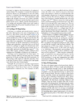
Figure 8. Schematic diagram of the developmental origins of Traditional skin substitutes either are made of natural
articular and growth plate cartilage [122]
8 International Journal of Bioprinting (2018)–Volume 4, Issue 1