
Page 23 - IJB-4-1
P. 23
Fan Liu, et al.
materials mimicking the composition of bone tissues (A) (B)
and the microenvironment of bony ECMs. For
example, in 2002, Ang et al. at National University
of Singapore printed a mesh hydroxyapatite-chitosan
structure as bone repair fillers [64] . In 2005, Seitz
et al. at Germany cooperated with Generis GmbH
(Augsburg, Germany) company developed a 3D
printed ceramic bone repair material [65] . In 2008,
Kouhi et al. at Australia Swinburne University of (C) (D)
Technology prepared a P400ABS plastic jawbone by
[66]
fused deposition manufacturing . In 2010, Smith et al.
at nScrypt company in Orlando produced a hard tissue
[67]
repair material using titanium and caprolactone . In the
same way, Lee et al. printed a porous calcium phosphate
cement/alginate scaffold by depositing a solution of
α-tricalcium phosphate-based powder and sodium alginate
[68]
in a calcium chloride bath . Comparing with the traditional
metal or polymethyl methacrylate (mechanical and semi- (E) (F)
mechanical) bone repair materials, most of the 3D printed
bone repair materials have two obvious characteristics: one
is made of biodegradable polymers, and the other is having
go-through channels or pores. The predefined channels in
the 3D printed construct are useful for nutrient supply and
metabolite elimination for the in-growth of osteoblasts [69–73] .
Some of the bone repair materials have showed good
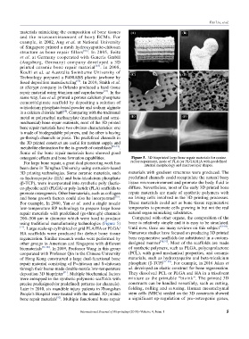
osteogenic effects and bone formation capabilities. Figure 5. 3D bioprinted large bone repair materials for canine
For large bone repair, a great deal pioneering work has radius repairment, made of PLA (or PLGA)/HA with predefined
internal morphology and macroscopic shapes.
been done in Tsinghua University using extrusion-based
3D printing technologies. Some ceramic materials, such materials with gradient structures were produced. The
as hydroxyapatite (HA) and beta-tricalcium phosphate predefined channels could recapitulate the natural bony
(β-TCP), were incorporated into synthetic poly (lactic- tissue microenvironment and promote the body fluid to
co-glycolic acid) (PLGA) or poly-lactide (PLA) scaffolds to diffuse. Nevertheless, most of the early 3D printed bone
promote osteogenesis. Other biomaterials, such as collagen repair materials are made of synthetic polymers with
and bone growth factors could also be incorporated [76] . no living cells involved in the 3D printing processes.
For example, In 2000, Yan et al. used a single nozzle These materials could act as bone tissue regenerative
low-temperature RP technology to prepare large bone temporaries to promote cells growing in but not the real
repair materials with predefined (go-through) channels natural organ mimicking substitutes.
200–500 μm in diameter which were hard to produce Compared with other organs, the composition of the
using traditional manufacturing technologies (Figure 5) bone is relatively simple and it is easy to be simulated.
[77] . Large scale-up cylindrical or grid PLA/HA or PLGA/ Until now, there are many reviews on this subject [83–89] .
HA scaffolds were produced for defect bone tissue Numerous studies have focused on producing 3D printed
regeneration. Similar research works were performed by bone regenerative scaffolds (or substitutes) in a custom-
other groups in American and Singapore with different designed manner [90,91] . Most of the scaffolds are made
biomaterials [78–80] . In 2009, Professor Wang in this group of synthetic polymers, such as PLGA, polycaprolactone
cooperated with Professor Qin in the Chinese University (PCL), with good mechanical properties, and ceramic
of Hong Kong constructed a large dual-functional bone materials, such as hydroxyapatite and beta-tricalcium
repair material consisting of P-chitosan and S-chitosan phosphate (β-TCP) [92–95] . For example, in 2016 Jakus et
through their home-made double-nozzle low-temperature al. developed an elastic construct for bone regeneration.
[81]
deposition 3D bioprinter . Multiple biochemical factors They dissolved PCL or PLGA and HA in a trisolvent
were entrapped in the synthetic polymeric scaffolds with mixture as the printable “bioink”. The printed 3D
precise predesigned (or predefined) patterns (or channels). constructs can be handled versatilely, such as cutting,
Later in 2010, six mandible injury patients in Zhongshan folding, rolling and suturing. Human mesenchymal
People’s Hospital were treated with the related 3D printed stem cells (MSCs) seeded on the 3D constructs showed
[82]
bone repair materials . Multiple functional bone repair a significant up-regulation of pro-osteogenic genes,
International Journal of Bioprinting (2018)–Volume 4, Issue 1 5