
Page 24 - IJB-4-1
P. 24
Progress in organ 3D bioprinting
collagen type I, osteocalcin, and osteopontin at day 28. laden constructs were submitted to a chemical crosslinking
When the 3D constructs were implanted in a macaque process to stabilize the structures and improve the
calvarial defect for 4 weeks, excellent new bone mechanical properties. Hepatocytes encapsulated in the
formation accompanying with the vascularization and gelatin-based hydrogels remained viable and produced
[96]
integration of surrounding tissue . At the same time, La hepatic ECMs during the 8 weeks’ in vitro culture. This
et al. reported a bone substitute that replicates the micro- is a great breakthrough in tissue engineering field which
and mineralized environment through printing PCL/ has encountered numerous bottleneck problems in organ
PLGA/TCP scaffolds, and then coating them with the manufacturing areas. Thus difficult problems, such as large
bone dECM (bdECM) that was extracted from bovine tissue formation and nutrient supply, have been solved
tibiae. The PCL/PLGA/TCP/bdECM scaffolds exhibited therefore. In 2007, a large scale-up vascularized liver
significantly enhanced osteogenic gene expression and tissue was first produced in the same group using another
calcium deposition. These experiments have further experiential CAD model [29,30] . From then, actual bioartifical
certified the bone regenerative effects of the PLGA/HA organ manufacturing has been put forward and developed
scaffolds which have been printed more than ten years very quickly. In 2009, a 3D printed complicated organ
ago in Professor Wang’s groups. with a whole fluent of endothelial layer covered the inner
3.2 Liver 3D Bioprinting channels of vascular network was produced [25–28] . It was
possible to observe that endothelial cells aligned inside the
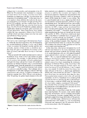
The liver is a vital visceral organ in the human body (Figure surface of the predefined channels. More than three cell
6). Unlike the structural organ bone, liver 3D bioprinting types formed functional tissues in a complex 3D construct.
has several bottleneck problems to solve: one of them This technique has advanced other researches at least ten
is how to construct the branched vascular and bile duct years in organ manufacturing areas [97,98] .
networks, another of them is how to distribute more At the same time, other groups throughout the world
than three cell types in a predefined 3D construct with a still devoted themselves in tissue engineered organ
high cell density and make them develop to functional dreams with their porous scaffolds. For example, Huang
[7]
tissues . et al. seeded hepatoma cells on a 3D printed branched
There are several CAD models have been used to construct vessel network which consists of avidin and biotin in
bioartificial livers. Some of the CAD models are made 2007 [99] . This is a typical traditional tissue engineering
of experience. For example, in 2004 professor Wang method to manufacture complex organs with a porous
and co-workers first assemble cell-laden gelatin-based scaffold. Later in 2013, Organovo company in American
hydrogels into large scale-up liver tissues with predefined printed a micro liver-tissue mimicking the techniques
structures (go-through channels) using a extrusion-based developed in Professor Wang’ group. According to the
3D printing system under the instruction of an experiential British New Scientist magazine website report, the
CAD model [16–19] . The predefined structures were printed micro-liver tissue, 0.5 mm in thickness, 4 mm in square
via a pressure-controlled syringe. This technique allows size, was created. To build the micro liver-tissue, two
the deposition of cell-laden hydrogels solutions with high main cell types of the liver, i.e. hepatocytes and hepatic
concentration and velocity. Cylindrical channels with stellate cells, were printed into 20 layers [100] . Cells in the
diameters ranging from 100 to 300 µm were produced. micro liver-tissue can survival for more than five days.
After 3D printing, the gelatin-based polymers in the cell- Neovascularization played a role in the cell survival
capabilities. In 2014, a bioartificial liver containing both
vascular and nervous networks has been produced layer-
by-layer using a combined MNRP under the instruction
of a much more complex experienced CAD model [101] .
The potential of this technology will eventually facilitate
the manufacture of bioartificial livers, and make the liver
3D bioprinting an impending reality.
Currently, there is a trend that to make the CAD models
from clinical patients. For instance, some current clinical
diagnostic technologies, such as computer tomography
(CT) and magnetic resonance imaging (MRI), have been
explored to acquire liver image information of the patients.
The CT and MRI image information are subsequently
transformed into CAD models (i.e. liver manufacturing
blueprints) and segregated into 2D horizontal slices to
provide instructions to the 3D bioprinters.
Figure 6. Schematic description the complex structure of the liver
6 International Journal of Bioprinting (2018)–Volume 4, Issue 1