
Page 22 - IJB-4-1
P. 22
Progress in organ 3D bioprinting
macromolecule (or polymer) formulation [43] . It is a and the physiological functionality realization of a
multi-layer procedure through the selective photo- supportive polymeric solution or hydrogel often need to
initiated curing reaction of a low-molecular weight pre- be addressed before the 3D bioprinting process.
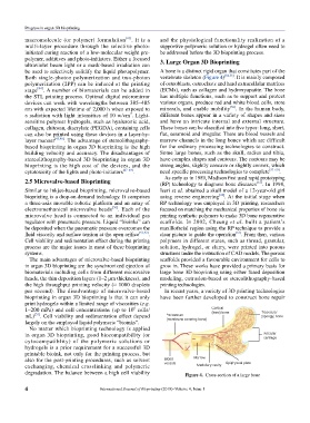
polymer, additives and photo-initiators. Either a focused 3. Large Organ 3D Bioprinting
ultraviolet beam light or a mask-based irradiation can
be used to selectively solidify the liquid photopolymer. A bone is a distinct rigid organ that constitutes part of the
Both single-photon polymerization and two-photon vertebrate skeleton (Figure 4) [54,55] . It is mainly composed
polymerization (2PP) can be induced at the printing of osteoblasts, osteoclasts and hard extracellular matrices
stage [44] . A number of biomaterials can be added in (ECMs), such as collagen and hydroxyapatite. The bone
the STL printing process. Optimal digital micromirror has multiple functions, such as to support and protect
devices can work with wavelengths between 385–405 various organs, produce red and white blood cells, store
nm with expected lifetime of 2,000 h when exposed to minerals, and enable mobility [56] . In the human body,
2
a radiation with light intensities of 10 w/cm . Light- different bones appear in a variety of shapes and sizes
sensitive polymer hydrogels, such as hyaluronic acid, and have an intricate internal and external structure.
collagen, chitosan, diacrylate (PEGDA), containing cells These bones can be classified into five types: long, short,
can also be printed using these devices in a layer-by- flat, sesamoid and irregular. There are blood vessels and
layer manner [45,46] . The advantage of stereolithography- marrow channels in the long bones which are difficult
based bioprinting in organ 3D bioprinting is the high for the ordinary processing technologies to construct.
building velocity and accuracy. The disadvantages of Some large bones, such as the skull, radius and tibia,
stereolithography-based 3D bioprinting in organ 3D have complex shapes and contours. The contours may be
bioprinting is the high cost of the devices, and the strong angles, slightly concave or slightly convex, which
cytotoxicity of the lights and photo-initiators [47–49] . need specific processing technologies to complete [57–60] .
As early as in 1989, Madison first used rapid prototyping
2.5 Microvalve-based Bioprinting (RP) technology to diagnose bone diseases [61] . In 1998,
Similar to inkjet-based bioprinting, microvalve-based Iseri et al. obtained a skull model of a 12-year-old girl
bioprinting is a drop-on-demand technology. It comprises using reverse engineering [62] . At the initial stage when
a three-axis movable robotic platform and an array of RP technology was employed in 3D printing, researchers
electromethanical microvalve heads [50] . Each of the focused on matching the mechanical properties of bone via
microvalve head is connected to an individual gas printing synthetic polymers to make 3D bone regenerative
regulator with pneumatic pressure. Liquid “bioinks” can scaffolds. In 2002, Cheung et al. built a patient’s
be deposited when the pneumatic pressure overcomes the maxillofacial region using the RP technique to provide a
[63]
fluid viscosity and surface tension at the open orifice [51,52] . clear picture to guide the operation . From then, various
Cell viability and sedimentation effect during the printing polymers in different states, such as thread, granular,
process are the major issues in most of these bioprinting solution, hydrogel, or slurry, were printed into porous
systems. structures under the instruction of CAD models. The porous
The main advantages of microvalve-based bioprinting scaffolds provided a favourable environment for cells to
in organ 3D bioprinting are the synchronized ejection of grow in. These works have provided a primary basis for
biomaterials including cells from different microvalve large bone 3D bioprinting using either fused deposition
heads, the thin deposition layers (1–2 µm thickness), and modeling, extrusion-based or stereolithography-based
the high throughput printing velocity (≈ 1000 droplets printing technologies.
per second). The disadvantage of microvalve-based In recent years, a variety of 3D printing technologies
bioprinting in organ 3D bioprinting is that it can only have been further developed to construct bone repair
print hydrogels within a limited range of viscosities (e.g.
1–200 mPa) and cell concentrations (up to 10 cells/
6
mL) [53] . Cell viability and sedimentation effect depend
largely on the employed liquid polymeric “bioinks”.
No matter which bioprinting technology is applied
in organ 3D bioprinting, good biocompatibility (or
cytocompatiblity) of the polymeric solutions or
hydrogels is a prior requirement for a successful 3D
printable bioink, not only for the printing process, but
also for the post-printing procedures, such as solvent
exchanging, chemical crosslinking and polymeric
degradation. The balance between a high cell viability Figure 4. Cross-section of a large bone
4 International Journal of Bioprinting (2018)–Volume 4, Issue 1