
Page 21 - IJB-4-1
P. 21
Fan Liu, et al.
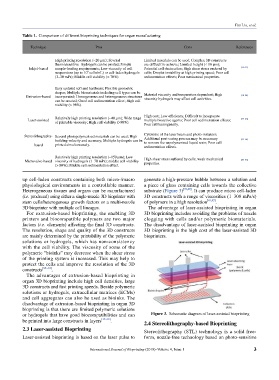
Table 1. Comparison of different bioprinting techniques for organ manufacturing
Technique Pros Cons References
High printing resolution (~20 µm); Several Limited materials can be used; Complex 3D constructs
thermosensitive hydrogels can be printed; Simple are difficult to achieve; Limited height (< 10 µm);
Inkjet-based sample-loading requirements; Low viscosity of cell Potential cell desiccation; High shear stress endured by [13–17]
6
suspensions (up to 10 cells/mL) or cell-laden hydrogels cells; Droplet instability at high printing speed; Poor cell
(3–30 mPs); Middle cell viability (> 70%). sedimentation effects; Poor mechanical properties.
Easy updated soft and hardware; Flexible geometric
shapes; Multiple biomaterials including cell types can be Material viscosity and temperature dependent; High
Extrusion-based incorporated; Homogeneous and heterogeneous structures viscosity hydrogels may affect cell activities. [18–24]
can be created; Good cell sedimentation effect; High cell
viability (> 98%).
Relatively high printing resolution (~40 µm); Wide range High cost; Low efficiency; Difficult to incorporate [39–42]
Laser-assisted of printable viscosity; High cell viability (>90%). multiple bioactive agents; Poor cell sedimentation effects;
Poor cell homogeneity.
Stereolithography- Several photopolymerized materials can be used; High Cytotoxic of the laser beam and photo-initiators;
building velocity and accuracy; Multiple hydrogels can be Additional post-curing process may be necessary [43–46]
based printed simultaneously. to remove the unpolymerized liquid resin; Poor cell
sedimentation effects.
Relatively high printing resolution (~150 µm); Low
Microvalve-based viscosity of hydrogels (1–70 mPs); middle cell viability High shear stress suffered by cells; weak mechanical [50–53]
properties.
(> 80%); Middle cell sedimentation effect.
up cell-laden constructs containing both micro-/macro generate a high-pressure bubble between a solution and
physiological environments in a controllable manner. a piece of glass containing cells towards the collective
Heterogeneous tissues and organs can be manufactured substrate (Figure 3) [39,40] . It can produce micro cell-laden
(i.e. produced) using either a single-nozzle 3D bioprinter with 3D constructs with a range of viscosities (1–300 mPa/s)
stem cells/heterogenous growth factors or a multi-nozzle of polymers in a high resolution [41,42] .
3D bioprinter with multiple cell lineages. The advantage of laser-assisted bioprinting in organ
For extrusion-based bioprinting, the enabling 3D 3D bioprinting includes avoiding the problems of nozzle
printers and biocompatible polymers are two major clogging with cells and/or polymeric biomaterials.
factors (i.e. elements) affecting the final 3D constructs. The disadvantage of laser-assisted bioprinting in organ
The resolution, shape and quality of the 3D constructs 3D bioprinting is the high cost of the laser-assisted 3D
are mainly determined by the printability of the polymeric bioprinters.
solutions or hydrogels, which has non-consistency
with the cell viability. The viscosity of some of the
polymeric “bioinks” may decrease when the shear stress
of the printing system is increased. This may help to
protect the cells and improve the resolution of the 3D
constructs [29–34] .
The advantages of extrusion-based bioprinting in
organ 3D bioprinting include high cell densities, large
3D constructs and fast printing speeds. Beside polymeric
solutions or hydrogels, extracellular matrices (ECMs)
and cell aggregates can also be used as bioinks. The
disadvantage of extrusion-based bioprinting in organ 3D
bioprinting is that there are limited polymeric solutions
or hydrogels that have good biocompatibilities and can Figure 3. Schematic diagram of laser-assisted bioprinting
be printed into large constructs in layers [35–38] . 2.4 Stereolithography-based Bioprinting
2.3 Laser-assisted Bioprinting Stereolithography (STL) technology is a solid free-
Laser-assisted bioprinting is based on the laser pulse to form, nozzle-free technology based on photo-sensitive
International Journal of Bioprinting (2018)–Volume 4, Issue 1 3