
Page 97 - IJB-4-1
P. 97
Pre-clinical evaluation of advanced nerve guide conduits using a novel 3D in vitro testing model
was connected to a high voltage supply (Genvolt UK) cultured at 37 °C in a humidified 95% air and 5% CO2
and the polymer-loaded syringe placed in the mount of atmosphere.
a programmable syringe pump (WPI Europe) and
constantly pumped through with a flow rate of 4
ml/hr. Polymer jet formation was achieved by a
voltage of 15 kV. The formed fibres were collected
on an earthed rotating aluminium collector (IKA
Works) wrapped in aluminium foil with a rotation
speed of 2000 rpm.
2.3 Characterisation of Microfibres
Gold coated electrospun fibre samples were imaged
using a XL-20 scanning electron microscope (SEM,
Koninklijke Philips N.V.) operating at 15 kV. On each
aluminium fibre sheet, three parallel arranged squares
were analysed regarding fibre diameter and density.
The fibre diameter was analysed using a ruler tool in
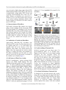
the SEM related XL software. The amount of fibres Figure 1. (A) Schematic workflow of the production of PCL
per 50 µm were counted and averaged over all microfibres using electrospinning, following the procedures of
[18]
images. Daud et al. , the analysis and threading procedure of microfibres
and the fabrication of PEG conduits by microstereolithography,
2.4 Combination of Conduit and Microfibers following the procedures of Pateman et al. [17] SEM
micrographs illustrating components of the nerve guide testing
Approximately 6000–7000 PCL microfibres were device; (B) A 5 mm long hollow PEG NGC, fabricated by
threaded per PEG conduit. The fibre sheets were cut to microstereolithography, and (C) PCL microfibres spun by
electrospinning. (D) Conduit and microfibres were combined
the required width and 1–2 cm of the fibres were into a final testing nerve guide device.
manually lifted off in the direction of the fibre
alignment. These fibres were twisted between the 2.6 F-actin Labelling of Neuronal Cells
fingers, where the twisted end was used in a similar NG108-15 neuronal cells were fixed with 3.7%
manner to a needle. PEG NGCs were threaded onto paraformaldehyde (PFA, v/v in distilled water,
PCL fibres (non-bunched end) like pearls on a chain Sigma) in whole NGCs for 3 h. For further staining
and fibre excess was cut with scissors. A schematic of procedures, the PCL microfibre filling was
the workflow can be found in Figure 1. carefully removed from the NGC by using forceps
and fixed on an objective slide. Cell membrane
2.5 Cell Culture in Whole Nerve Guides
permeabilisation was conducted with 0.1% Triton
NG108–15 neuroblastoma × glioma rat/mouse hybrid X-100 (w/v, Sigma) in phosphate buffered saline
neuronal cells (Public Health England, UK) were (PBS, Thermo Scientific) for one hour. Cytoskeleton’s
cultured in whole nerve guides with a cell concentration F-actin was visualised by using phalloidin
of 6 × 10 cells in proliferation medium, containing of conjugated to tetramethylrhodamine (TRITC) (v/v
5
Dulbecco’s Modified Eagle Medium (DMEM, Sigma), 1:1000 dilution in PBS, Sigma), and cell nuclei
10% foetal bovine serum (FBS, v/v, Biosera), 0.25 were labelled with 4',6-diamidino-2-phenylindole
mg/mL amphotericin (Sigma), 200 mM L-glutamine (DAPI, 300 nM, Sigma) for 1 h at room temperature.
(Sigma), 100 units/mL penicillin and 100 mg/mL 2.7 Live/Dead Cell Staining of Neuronal Cells
streptomycin (Sigma) for 4 days. Cells were seeded with
15 µL of cell suspension by directly pipetting on top of To distinguish live and dead neuronal cells visually,
the fibres in the conduits. The NGCs were transferred to living cells were stained green by using 0.02% Syto 9
the designed culturing setup in a 6-well plate and cells (v/v, Thermo Fisher Scientific) and dead cells red by
were allowed to attach at 37 °C for 30 minutes before using 0.03% propidium iodide (v/v, Thermo Fisher
wells were filled with proliferation medium. Neuronal Scientific) in serum-free medium (proliferation
NG108–15 cells were used between passage 14–20 and medium deprived from serum). Cells were incubated
International Journal of Bioprinting (2018)–Volume 4, Issue 1 3