
Page 98 - IJB-4-1
P. 98
Mehri Behbehani, et al.
in the staining solution for 60 min at 37 °C. Confocal on a glass microscope slide and imaged in PBS using
imaging was conducted in PBS immediately after a 10× magnification ZeissW Plan Achromat water-
staining (details below). dipping objective lens. For imaging FITC- and SYTO
9-labelled samples incident and excitation wavelengths
2.8 Dorsal Root Ganglion Isolation and Culture of λex = 488 nm/λem = 525 nm were used, and
Male Wistar rats aged 10–12 weeks were sacrificed by wavelengths of λex = 543 nm/λem = 576 nm to image
cervical dislocation (schedule I procedure, UK Home Texas Red, TRITC and propidium iodide-labelled
Office). Rats were skinned and the spine was removed. samples. Cell nuclei were visualized at λex = 780
DRGs were extracted after the spine was cut open, nm/λem = 480 nm. Images were stitched together and
dorsal side facing up, and the spinal cord and analysed using Zeiss LSM Image Browser software
meninges were removed. The nerve roots of each and Image J 1.49 (National Institute of Health, USA).
DRG were trimmed and explant DRG bodies placed
on top of the nerve guides (one DRG body per
conduit), which were held in place by the described
setup. DRGs were incubated at 37 °C for 15 min to
allow attachment. Afterwards, samples were fully
covered with proliferation medium and incubated at
37 °C in a humidified 95% air and 5% CO 2 atmosphere
for 21 days.
2.9 βIII-tubulin and S100β Labelling of Dorsal
Root Ganglia
In order to reveal neuron-specific protein βIII-tubulin
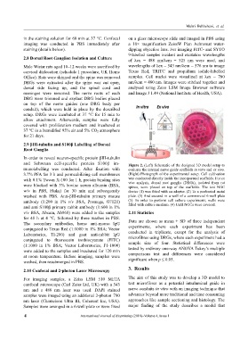
and Schwann cell-specific protein S100β im- Figure 2. (Left) Schematic of the designed 3D model setup to
munolabelling was conducted. After fixation with evaluate the internal nerve guide scaffolds in vitro and ex vivo.
3.7% PFA for 3 h and permeabilising cell membranes (Right) Photograph of the experimental setup. Cell cultivation
with 0.1% Tween X-100 for 1 h, protein binding sites was conducted directly inside the incorporated scaffolds. For ex
vivo analysis, dorsal root ganglia (DRGs), isolated from rat
were blocked with 3% bovine serum albumin (BSA, spines, were placed on top of the scaffolds. The test NGC
w/v in PBS, Fluka) for 30 min and subsequently device (1) was fitted with an adapter. (2) To a perforated metal
washed with PBS. Anti-βIII-tubulin primary mouse plate. (3) And secured in a well of a commercial 6-well plate
antibody (1:200 in 1% v/v BSA, Promega, G7121) (5). In order to perform cell culture experiments, wells were
filled with culture medium. (4) Until NGCs were covered.
and anti S100β primary rabbit antibody (1:600 in 1%
v/v BSA, Abcam, Ab868) were added to the samples 2.11 Statistics
for 48 h at 4 °C, followed by three washes in PBS.
The secondary antibodies, horse anti-mouse lgG Data are shown as mean ± SD of three independent
conjugated to Texas Red (1:1000 in 1% BSA; Vector experiments, where each experiment has been
Laboratories, TI-200) and goat anti-rabbit lgG conducted in triplicate, except for the analysis of
conjugated to fluorescein isothiocyanate (FITC) microfibres using DRGs, where each experiment had a
(1:1000 in 1% BSA; Vector Laboratories, F1-1000) sample size of four. Statistical differences were
were added to the samples and incubated for 120 min tested by ordinary one-way ANOVA Tukey’s multiple
at room temperature. Before imaging, samples were comparisons test and differences were considered
washed, then resubmerged in PBS. significant when p ≤ 0.05.
3. Results
2.10 Confocal and 2-photon Laser Microscopy
For imaging samples, a Zeiss LSM 510 META The aim of this study was to develop a 3D model to
confocal microscope (Carl Zeiss Ltd, UK) with a 543 test microfibres as a potential intraluminal guide in
nm and a 488 nm laser was used. DAPI stained nerve conduits in vitro with an imaging technique that
samples were imaged using an additional 2-photon 780 advances beyond more traditional and time consuming
nm laser (Chameleon Ultra III, Coherent Inc, USA). approaches like sample sectioning and histology. The
Samples were arranged in a 6-well plate or were fixed major finding of the study describes a model that
4 International Journal of Bioprinting (2018)–Volume 4, Issue 1