
Page 100 - IJB-4-2
P. 100
Optimized vascular network by stereolithography for tissue engineered skin
released cytotoxic compounds during the first two days
of elution: revealing moderate to strong cytotoxicity at
day 1 and weak to moderate cytotoxicity at day 2. The
®
material Irgacure 184 did not cause cytotoxicity at day
1 and a weak moderate at day 2.
®
BLI with Irgacure 184 showed no to low cytotoxicity
in both live/dead and WST-1 assay. For the sample BLI
®
with Irgacure 2959, very high cytotoxicity was found
in the live/dead assay. Although for the sample BLI with
®
Irgacure 369 no or only low cytotoxicity was observed
in the live/dead assay, a release of cytotoxic compounds
was detected at the beginning of elution. Besides,
the data from staining could only poorly be assessed
because these samples showed a very high background
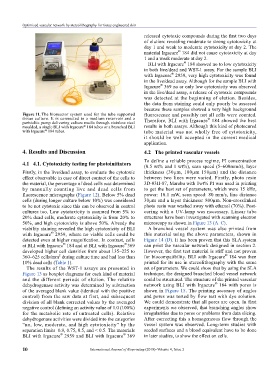
Figure 11. The bioreactor system used for the tube supported fluorescence and possibly not all cells were counted.
tissue culture. It is connected to a medium reservoir and a ®
peristaltic pump delivering culture media through stainless steel Therefore, BLI with Irgacure 184 showed the best
moulded, a single BLI with Irgacure 184 tubes or a branched BLI results in both assays. Although this kind of photocured
®
with Irgacure 184 tubes. tube material was not wholly free of cytotoxicity,
®
it should be well accepted in the current medical
application.
4. Results and Discussion 4.2 The printed vascular vessels
To define a reliable process regime, PI concentration
4.1 4.1. Cytotoxicity testing for photoinitiators (0.5 wt% and 1 wt%), scan speed (5–600mm/s), layer
Firstly, in the live/dead assay, to evaluate the cytotoxic thickness (30µm, 100µm 150µm) and the distance
effect observable in case of direct contact of the cells to between two lines were varied. Firstly, photo resin
the material, the percentage of dead cells was determined 3D-03H-87, Marabu with 1wt% PI was used in printing
by manually counting live and dead cells from to get the best set of parameters, which were 15 kHz,
fluorescence micrographs (Figure 12). Below 5% dead power: 10.1 mW, scan speed: 80 mm/s, line distance
cells (during longer culture below 10%) was considered 30µm and a layer thickness: 800µm. Non-crosslinked
to be not cytotoxic since this can be observed in control photo resin was washed away with ethanol (70%). Post-
cultures too. Low cytotoxicity is assumed from 5% to curing with a UV-lamp was necessary. Linear tube
20% dead cells, moderate cytotoxicity is from 20% to structures have been investigated with scanning electron
50%, and high cytotoxicity is above 50%. Already the microscopy as shown in Figure 13 (A–C).
viability staining revealed the high cytotoxicity of BLI A branched vessel system was also printed from
®
with Irgacure 2959, where no viable cells could be this material using the above parameters, shown in
detected even at higher magnification. In contrast, cells Figure 14 (D). It has been proven that this SLA system
®
®
at BLI with Irgacure 184 and at BLI with Irgacure 369 can print the vascular network designed in section 2.
developed higher cell densities from about 135–225 to However, the first test material is stiff and not proven
®
2
360–625 cells/mm during culture time and had less than for biocompatibility. BLI with Irgacure 184 was then
10% dead cells (Table 1). printed for its use in stereolithography with the same
The results of the WST-1 assays are presented in set of parameters. We could show that by using the SLA
Figure 13 as boxplot diagrams for each kind of material technique, the designed branched blood vessel network
and the different periods of elution. The relative could be structured. The structure of the printed vascular
®
dehydrogenase activity was determined by subtraction network using BLI with Irgacure 184 with pores is
of the averaged blank value (identical with the positive shown in Figure 15. The printing accuracy of angles
control) from the raw data at first, and subsequent and pores was tested by flow test with dye solution.
division of all blank corrected values by the averaged We could demonstrate that all pores are open. In first
negative control (defining an activity value of 1.0 (100%) experiments we observed, that branching angles show
for the metabolic rate of untreated cells). Relative irregularities due to pores or problems from data slicing.
dehydrogenase activities were divided into the categories After correcting this a homogeneous flow through the
“no, low, moderate, and high cytotoxicity” by the vessel system was observed. Long-term studies with
separation limits 0.9, 0.75, 0.5, and < 0.5. The materials seeded surfaces and a blood equivalent have to be done
®
®
BLI with Irgacure 2959 and BLI with Irgacure 369 in later studies, to show the effect on cells.
10 International Journal of Bioprinting (2018)–Volume 4, Issue 2