
Page 98 - IJB-4-2
P. 98
Optimized vascular network by stereolithography for tissue engineered skin
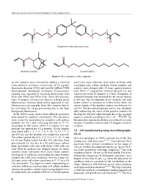
Figure 9. Three monomers in this composite
on the samples were stained by adding a twofold activities) were obtained from wells without cells
concentrated solution consisting of 30 µg/mL containing only culture medium. Eluate samples and
fluorescein diacetate (FDA) and twofold GelRed (VWR controls were changed after 24 hours against medium
®
International, Darmstadt, Germany). Fluorescence with WST-1 reagent but without phenol red and
staining was captured by Axiotech microscope with cultured for about 20 minutes to 1 hour. Formation of
filter sets FS09 and FS14 (Carl Zeiss Microscopy, coloured formazan was measured by the optical density
Jena, Germany). Living cells showed a bright green at 450 nm. The development of dye intensity was kept
fluorescence, whereas dead nuclei appeared in red. under control to measure at a time point, when the
Fluorescence micrographs from BLI samples had to optical density of the negative control was between 0.2
be contrasted by image processing due to the high and 0.6. Relative dehydrogenase activity was calculated
background fluorescence. after subtracting the mean of the positive controls by
In the WST-1 assay, eluates from different specimens dividing the obtained values through that of the averaged
were tested for cytotoxic components. The specimens negative controls according to . By
were eluted by incubation in complete cell culture this procedure measured activities were placed on a scale
medium for 24 h and following periods at 37 °C. between 0 (positive control) and 1.0 (negative control)
According to ISO norm 10993-5 a volume of 1 mL or above.
medium was applied per 0.2 g material. Eluate samples
were taken after 1, 2, 3, 6, 7, 8, 9, 10, 13, 15, 17, 21 3.2 AM manufacturing using stereolithography
and 24 days and the medium was completely renewed. (SLA)
The elution period day 0–1, 1–2, 2–3, 6–7, 10–13 and SLA was developed in 1980’s and was one of the first
21–24 was finally tested. For this 3T3 cells have been commercial AM processes [41,42] . Conventional SLA
pre-cultured for one day in a 96 well tissue culture machines have vertical resolutions in the range of
plate inoculated with care with about 8,000 cells per 150 µm. Further developments known as “micro SLA”
well. Then the medium was changed against the eluate can create geometries with high com plexity [43] and with
samples (four replicates from each eluate sample resolutions below 150 µm in all three spatial directions.
resulting in 12 replicates representing the same material Behind the background of biomimetic structures, layer
sample) and cells were incubated with the eluates for heights of less than 10 µm, e.g. allow the replication of
24 hours under cell culture conditions at 37 °C in a 5% capillaries that are essential for the metabolism in the
CO atmosphere. The negative control (representing no tissue. Alternative AM methods are not able to produce
2
cytotoxic influence) received pure cell culture medium such high-resolution structures [13,44,45]. The high resolution
whereas the positive control (representing highest level of AM cell scaffolds or membranes enables targeted cell
of cytotoxicity and complete inhibition of dehydrogenase alignment, cell growth and cell interaction [44,46] . The SLA
8 International Journal of Bioprinting (2018)–Volume 4, Issue 2