
Page 117 - IJB-4-2
P. 117
Physical stimulations and their osteogenesis-inducing mechanisms
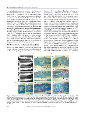
current stimulation could promote bone formation Azuma et al. [131] investigated the effects of ultrasonic
2
of rabbit posterolateral fusion model. Park et al. [129] stimulation (30 mW/cm , 20 min/day) on fracture
investigated the effects of electric stimulation of 1h/day healing in the different duration (days 1–8, 9–16, 17–24
for 4 weeks on 3 mm gapped osteotomies in mid-tibial and 1–24). The radiography and histological results
of rabbit models, the two electrodes were placed on the demonstrated that the low intensity pulsed ultrasound
above patellar tendon and lateral thigh, respectively. The could accelerate fracture healing at each treatment period,
results showed that the callus area and mineral content and the 1–24 days group was more effective than other
were 27% and 31% higher than control osteotomies, treated groups (Figure 6). Moreover, the mechanical
respectively, and the biomechanical properties were torsion properties of treated femurs were significantly
significantly higher than control group. It indicated that higher than nontreated femurs, and the properties in the
the electric stimulation could increase the mineralization 1–24 days group were the highest. Takikawa et al. [132]
and callus development of the bone healing regions, established nonunion model of tibia fracture in rat, and
2
thereby enhancing the biomechanical properties. utilized low intensity pulsed ultrasound (30 mW/cm ) to
Chen et al. [130] evaluated the changes in bone mineral treat the fracture sites. They found that the healing rates
density of fifteen males with spinal cord injury after the of tibia samples were 30.8% and 50% after treated for 4
intervention of functional electric stimulation 6 months. weeks and 6 weeks, respectively, while the samples in
The results showed that the bone mineral density control group were not healing. Nolte et al. [115] studied
was increased significantly, whereas the effect would the bioeffect of low intensity pulsed ultrasound (20 min/
disappear when the stimulation was removed. day) on the fracture nonunion sites of 29 patients. The
results showed that 86% of patients obtained complete
4.3 In vivo Studies of Mechanical Stimulation healing after 22 weeks. Fritton et al. [133] investigated the
Mechanical stimulation can accelerate the bone repair skeletal responsed to compressive loads by applying
process and induce healing of nonunions, which depends controlled cyclic axial load on mouse tibia and analyzed
on the intensity, frequency and duration of loading. the bone mineral content of loaded and unloaded
A C D
A1 C1 D1
A2
A3
C2 D2
A4
A5
B C3 D3
B1 B2
Figure 6. Effects of low intensity pulsed ultrasound on fracture healing in the different duration. (A) Radiography of treated femur (A2-
A5, treatment duration at days 1–8, 9–16, 17–24 and 1–24, respectively) and nontreated femur (A1) at day 25 after fracture. The treated
groups had better bone healing than control group. (B–D) Histological analyses of low intensity pulsed ultrasound treatment on fracture
healing at different duration. At day 9, early endochondral ossification in treated femur (B2) was greater than in the control (B1). At day
17, endochondral ossification and remodeling in the control femur (C1) were less than treated femur 16 days (days 1–16, C2) and 8 days
(days 9–16, C3). At day 25, bone bridging in the control femur (D1) was less than treated 24 days (days 1–24, D2) and 8 days (days 17–
24, D3) [131] .
10 International Journal of Bioprinting (2018)–Volume 4, Issue 2