
Page 114 - IJB-4-2
P. 114
Shuai C, et al.
osteoblasts which was critical for mechanosensation and customized porous scaffolds present a great challenge
intracellular signal transduction. And the actin filaments for manufacturing process. 3D printing is one of the
rapidly reorganized into thick parallel bundles of fibres, advanced manufacturing technologies which fabricate
and the fibre formation was induced by shear stress objects directly from the given computer-aided design
loading 0-90 min whereas the cytoskeleton was disrupted model via layer by layer printing. It can fabricate
over loading 90 min (Figure 3B). Besides, fluid shear the interconnected internal porous structure and the
stress could produce bioeffects to cells which seeded on customized external shape of bone scaffolds. Moreover,
3D printed bone scaffolds [95,96] . Stiehler et al. studied the bone scaffolds require excellent biocompatibility to
effect of fluid shear stress on human mesenchymal stem encourage cell adhesion and migration [99] . Bioceramics
cells cultured on porous poly(D,L-lactide-co-glycolide) (such as hydroxyapatite, bioactive glass, etc.) and
scaffolds, and the results showed that the fluid shear biopolymers (such as polycaprolactone, polylactide,
stress markedly enhanced alkaline phosphatase activity, etc.) are suitable materials for the fabrication of bone
2+
[95]
increased Ca content and promoted cells growth . scaffolds owing to their good biological properties [100–102] .
3.4 Physical Stimulations on Artificial Bone Magnetic materials (such as Fe O , γ-Fe O , etc.)
4
3
2
3
and conductive materials (such as carbon nanotube,
In terms of bone defects repair, bone scaffolds need to graphene, etc.) are incorporated in bioceramics and/
possess interconnected internal porous structures that or biopolymers to enhance the biological and physical
provide channels for the adhesion and migration of bone properties of scaffolds [103–106] . Zhang et al incorporated
cells, the transmission of nutrients, and the growth of Fe O nanoparticles into polycaprolactone and meso-
3
4
bone tissue [97] . Meanwhile, bone scaffolds also need porous bioactive glass composites, and found that the
to possess customized external geometries that can 3D printed composite scaffold significantly stimulated
exactly match bone defects, which is beneficial for the cells proliferation and differentiation [107] . Therefore, the
structural and functional remodeling of bone [98] . The bone scaffold fabricated via 3D printing technology with
A B
A1 A2 B1 B2
A3 A4 B3 B4
A5 A6 B5 B6
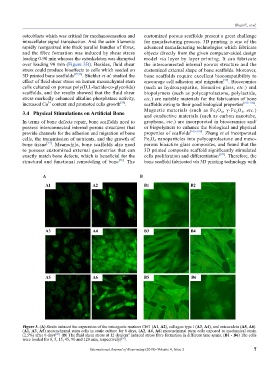
Figure 3. (A) Strain induced the expression of the osteogenic markers Cbf1 (A1, A2), collagen type I (A3, A4), and osteocalcin (A5, A6).
(A1, A3, A5) mesenchymal stem cells in static culture for 6 days, (A2, A4, A6) mesenchymal stem cells exposed to mechanical strain
(2.5%) after 6 days . (B) The fluid shear stress at 12 dyn/cm induced stress fibre formation in different time spans. (B1 - B6) The cells
2
[90]
were loaded for 0, 5, 15, 45, 90 and 120 min, respectively) .
[94]
International Journal of Bioprinting (2018)–Volume 4, Issue 2 7