
Page 71 - IJB-4-2
P. 71
Novel ultrashort self-assembling peptide bioinks for 3D culture of muscle myoblast cells
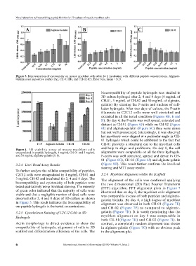
Figure 3. Representation of cytotoxicity on mouse myoblast cells after 24 h incubation with different peptide concentrations. Alginate-
Gelatin used as positive control (A), CH-01 (B), and CH-02 (C). Error bars, mean ± S.D.
biocompatibility of peptide hydrogels was studied in
3D culture hydrogel after 2, 4 and 8 days (4 mg/mL of
CH-01, 3 mg/mL of CH-02 and 30 mg/mL of alginate-
gelatin) By staining the F-actin and nucleus of cell-
laden hydrogels. After two days of culture, the F-actin
filaments in C2C12 cells were well stretched and
extended in all the tested condition (Figures 6B, E and
H). By day 4, the F-actin was well spread, extended and
distinct in CH-01 (Figure 6F) while on CH-02 (Figure
6I) and alginate-gelatin (Figure 6C) they were dense
but not well pronounced. Interestingly, it was observed
hat myoblasts were aligned at a particular angle in CH-
01 hydrogels which could be attributed to the fact that
CH-01 provides a structural cue to the myoblast cells
and help to align and proliferate. On day 8, the cell
Figure 4. 3D viability assay of mouse myoblast cells
encapsulated in peptide hydrogels, 4 mg/mL CH-01 and 3 mg/mL alignments were comparable on all the three hydrogels.
and 30 mg/mL alginate-gelatin (1:1). F-actin was well stretched, spread and dense in CH-
01 (Figure 6G), CH-02 (Figure 6J) and alginate-gelatin
3.2.4 Live/ Dead Assay Results (Figure 6D). This result further confirms the live/dead
staining and MTT assay results.
To further analyze the cellular compatibility of peptides,
C2C12 cells were encapsulated in 4 mg/mL CH-01 and 3.2.6 Myoblast Alignment within the Scaffold
3 mg/mL CH-02 and incubated for 2, 4 and 8 days. The The alignment of the cells was confirmed applying
biocompatibility and cytotoxicity of both peptides were the two dimensional (2D) Fast Fourier Transform
tested qualitatively using live/dead staining. The intensity (FFT) algorithm. FFT alignment plots in Figure 7
of green color indicated that the majority of cells were illustrated that on day 2, the myoblast cells alignment
viable and that a negligible number of dead cells were was comparable in case of both peptides and alginate-
observed after 2, 4 and 8 days of 3D-culture as shown gelatin bioinks. By day 4, a high degree of myoblast
in Figure 5. This result indicates the biocompatibility of alignment was observed in both CH-01 (Figure 7E)
our peptide hydrogels in the tested concentrations. and CH-02 (Figure 7F) as compared to alginate-
3.2.5 Cytoskeleton Staining of C2C12 Cells in 3D gelatin (Figure 7D). It is worth mentioning that the
Hydrogels myoblast alignment on day 8 was comparable in
both CH-01(Figure 7H) and CH-02 (Figure 7I). In
Actin morphology is direct evidence to show the contrast, a completely random alignment was shown
compatibility of hydrogels, alignment of cells in 3D in alginate-gelatin (Figure 7G) with no obvious peak
scaffold and differentiation efficiency of the cells. The in the alignment plot.
6 International Journal of Bioprinting (2018)–Volume 4, Issue 2