
Page 186 - IJB-10-4
P. 186
International Journal of Bioprinting Effects of structure on the interbody cage
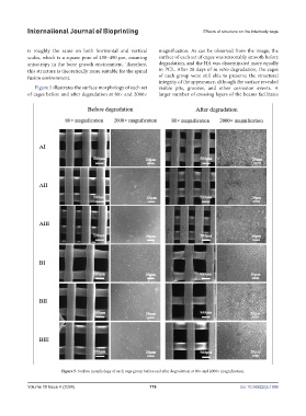
is roughly the same on both horizontal and vertical magnification. As can be observed from the image, the
scales, which is a square pore of 450–490 μm, ensuring surface of each set of cages was reasonably smooth before
anisotropy in the bone growth environment. Therefore, degradation, and the HA was disseminated more equally
this structure is theoretically more suitable for the spinal in PCL. After 28 days of in vitro degradation, the cages
fusion environment. of each group were still able to preserve the structural
integrity of the appearance, although the surface revealed
Figure 5 illustrates the surface morphology of each set visible pits, grooves, and other corrosion events. A
of cages before and after degradation at 80× and 2000× larger number of crossing layers of the beams facilitates
Figure 5. Surface morphology of each cage group before and after degradation at 80× and 2000× magnification.
Volume 10 Issue 4 (2024) 178 doi: 10.36922/ijb.1996