
Page 187 - IJB-10-4
P. 187
International Journal of Bioprinting Effects of structure on the interbody cage
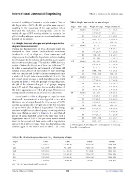
increased visibility of corrosion on the surface. Due to Table 3. Weight loss rates for each set of cages
the degradation of PCL, the HA particles were exposed, Cages Time (day) Weight loss (mg) Weight loss rate (%)
resulting in the roughness of the cage surface, which
facilitates the induction of osteogenesis. Due to the AI Control 0.65 ± 0.21 0.11 ± 0.03
weekly change of SPF soaking solution to maintain the 7 6.07 ± 0.49 1 ± 0.14
pH of the degrading environment, no mineralization was 14 7.53 ± 1.27 1.24 ± 0.21
noticed on the surface. 21 11.85 ± 2.33 1.76 ± 0.32
3.3. Weight loss rate of cages and pH changes in the 28 19.1 ± 1.27 3.12 ± 0.1
degradation environment AII Control 0.4 ± 0 0.07 ± 0
During the decomposition of PCL, chemical bonds are 7 6.1 ± 0.44 1.06 ± 0.11
disrupted to form certain small-molecule monomers 14 7.73 ± 0.84 1.36 ± 0.32
(6-ethanolic acid) or oligomers. These monomers and 21 9.83 ± 1.5 1.83 ± 0.13
oligomers are dissolved in the degradation solution, leading 28 20 ± 3.96 3.42 ± 0.15
to pH changes in the solution and contributing to quality
decline of the complete cage. The addition of HA also has a AIII Control 0.4 ± 0.14 0.07 ± 0.02
40
certain effect on the alterations of these two indicators. 19,20 . 7 6.67 ± 0.32 1.12 ± 0.05
In order to reconstruct the environment of dynamic pH 14 8.1 ± 1.2 1.37 ± 0.24
balance in vivo, the pH of the solution in each centrifuge 21 10.55 ± 0.92 1.83 ± 0.23
tube was checked and the SBF solution was replaced once 28 19.95 ± 4.88 3.43 ± 0.3
a week, and the pH value was controlled at 7.4 ± 0.3. The BI Control 0.25 ± 0.15 0.05 ± 0.02
pH of each group of cages at each degradation time point 7 5.77 ± 0.06 1.17 ± 0.06
is given in Table 2. With the passage of degrading time,
the pH of the solutions dropped in all groups, ranging 14 7.63 ± 0.74 1.53 ± 0.15
from 0.23 to 0.45. This suggests that some degradation of 21 9.6 ± 1.04 1.88 ± 0.26
the fusion apparatus occurred in all groups. However, no 28 16.8 ± 1.98 3.28 ± 0.28
strong association with structural traits was discovered. BII Control 0.3 ± 0.17 0.06 ± 0.01
As indicated in Table 3, all groups of cages lost mass 7 6.03 ± 0.31 1.18 ± 0.02
slowly with the extension of in vitro degradation time, with 14 7.73 ± 0.21 1.58 ± 0.12
the lowest rate of weight loss of the AI structure of 3.12% 21 9.93 ± 0.87 1.96 ± 0.12
and the maximum rate of weight loss of the BIII structure 28 21.35 ± 1.91 3.96 ± 0.2
of up to 4.49% after 28 days of degradation. The change BIII Control 0.45 ± 0.26 0.09 ± 0.05
curves of weight loss rate of each group versus degradation
time were plotted, as shown in Figure 6. It is seen that all 7 6.17 ± 0.12 1.29 ± 0.06
groups of cages degraded faster in the first week with a 14 8.17 ± 0.12 1.64 ± 0.09
degradation rate of 0.89–1.29% per week, which slowed 21 10.97 ± 0.32 2.24 ± 0.19
down in the second and third weeks with a degradation 28 22.95 ± 0.35 4.49 ± 0.15
rate of 0.36–0.48% per week. Then the degradation rate A: 60% filling rate, B: 40% filling rate; I: 1 crossing layer, II: 2 crossing
climbed again in the fourth week to about 1.00–2.25% layers, III: 4 crossing layers.
Table 2. The pH of each degradation node (pH ) of each group of cages
t
Cages 7 days 14 days 21 days 28 days
AI 7.12 ± 0.11 7.04 ± 0.06 7.05 ± 0.05 7.04 ± 0.09
AII 7.11 ± 0.07 7.1 ± 0.05 7.12 ± 0.08 7.11 ± 0.08
AIII 7.09 ± 0.04 7.11 ± 0.05 7.1 ± 0.06 7.11 ± 0.11
BI 7.19 ± 0.04 7.04 ± 0.08 7.03 ± 0.07 7.03 ± 0.06
BII 7.17 ± 0.05 7.09 ± 0.05 6.96 ± 0.06 7.03 ± 0.08
BIII 7.18 ± 0.09 7.04 ± 0.09 7 ± 0.09 7.02 ± 0.07
pH = 7.41. SBF was replaced weekly to reset pH.
0
Volume 10 Issue 4 (2024) 179 doi: 10.36922/ijb.1996