
Page 367 - IJB-10-4
P. 367
International Journal of Bioprinting 3D-printed PEEK in cranioplasty
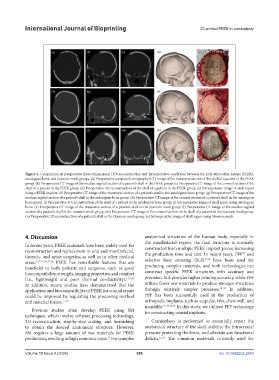
Figure 4. Comparison of preoperative three-dimensional (3D) reconstruction and intraoperative conditions between the poly-ether-ether-ketone (PEEK),
autologous bone, and titanium mesh groups. (a) Preoperative computed tomography (CT) image of the transverse section of the skull of a patient in the PEEK
group. (b) Preoperative CT image of the median sagittal section of a patient’s skull in the PEEK group. (c) Preoperative CT image of the coronal section of the
skull of a patient in the PEEK group. (d) Preoperative 3D reconstruction of the skull of a patient in the PEEK group. (e) Intraoperative image of skull repair
using a PEEK implant. (f) Preoperative CT image of the transverse section of a patient’s skull in the autologous bone group. (g) Preoperative CT image of the
median sagittal section of a patient’s skull in the autologous bone group. (h) Preoperative CT image of the coronal section of a patient’s skull in the autologous
bone group. (i) Preoperative 3D reconstruction of the skull of a patient in the autologous bone group. (j) Intraoperative image of skull repair using autologous
bone. (k) Preoperative CT image of the transverse section of a patient’s skull in the titanium mesh group. (l) Preoperative CT image of the median sagittal
section of a patient’s skull in the titanium mesh group. (m) Preoperative CT image of the coronal section of the skull of a patient in the titanium mesh group.
(n) Preoperative 3D reconstruction of a patient’s skull in the titanium mesh group. (o) Intraoperative image of skull repair using titanium mesh.
4. Discussion anatomical structures of the human body, especially in
the maxillofacial region, the final structure is normally
In recent years, PEEK materials have been widely used for constructed from multiple PEEK implant pieces, increasing
reconstruction and replacement in oral and maxillofacial, the production time and cost. In recent years, FFF and
32
thoracic, and spine surgeries, as well as in other medical 33,34
areas. 5,17,19,20,27,28 PEEK has remarkable features that are selective laser sintering (SLS) have been used for
beneficial to both patients and surgeons, such as good producing complex materials, and both technologies can
biocompatibility, strength, imaging properties, and comfort construct specific PEEK structures with accuracy and
(i.e., lightweight and poor thermal conductivity). 1,29,30 precision. SLS provides higher printing accuracy, while FFF
In addition, recent studies have demonstrated that the utilizes fewer raw materials to produce stronger structures
application and biocompatibility of PEEK for cranial repair through relatively simpler processes. 35-38 In addition,
could be improved by regulating the processing method FFF has been successfully used in the production of
and material fusion. 11,29 orthopedic implants, such as scapulae, ribs, chest wall, and
mandible. 17,19,20,39 In this study, we utilized FFF technology
Previous studies often develop PEEK using SM for constructing cranial implants.
techniques, which involve software processing technology,
3D reconstruction, step-by-step cutting, and burnishing Cranioplasty is performed to essentially repair the
to obtain the desired anatomical structure. However, anatomical structure of the skull, stabilize the intracranial
SM requires a large amount of raw materials for PEEK pressure protecting the brain, and alleviate any functional
production, resulting in high economic costs. For complex deficits. 12,13 The common materials currently used for
31
Volume 10 Issue 4 (2024) 359 doi: 10.36922/ijb.2583