
Page 368 - IJB-10-4
P. 368
International Journal of Bioprinting 3D-printed PEEK in cranioplasty
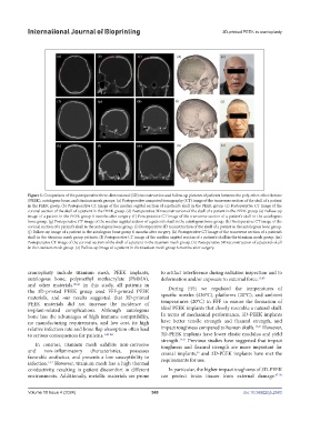
Figure 5. Comparison of the postoperative three-dimensional (3D) reconstruction and follow-up pictures of patients between the poly-ether-ether-ketone
(PEEK), autologous bone, and titanium mesh groups. (a) Postoperative computed tomography (CT) image of the transverse section of the skull of a patient
in the PEEK group. (b) Postoperative CT image of the median sagittal section of a patient’s skull in the PEEK group. (c) Postoperative CT image of the
coronal section of the skull of a patient in the PEEK group. (d) Postoperative 3D reconstruction of the skull of a patient in the PEEK group. (e) Follow-up
image of a patient in the PEEK group 6 months after surgery. (f) Postoperative CT image of the transverse section of a patient’s skull in the autologous
bone group. (g) Postoperative CT image of the median sagittal section of a patient’s skull in the autologous bone group. (h) Postoperative CT image of the
coronal section of a patient’s skull in the autologous bone group. (i) Postoperative 3D reconstruction of the skull of a patient in the autologous bone group.
(j) Follow-up image of a patient in the autologous bone group 6 months after surgery. (k) Postoperative CT image of the transverse section of a patient’s
skull in the titanium mesh group patients. (l) Postoperative CT image of the median sagittal section of a patient’s skull in the titanium mesh group. (m)
Postoperative CT image of the coronal section of the skull of a patient in the titanium mesh group. (n) Postoperative 3D reconstruction of a patient’s skull
in the titanium mesh group. (o) Follow-up image of a patient in the titanium mesh group 6 months after surgery.
cranioplasty include titanium mesh, PEEK implants, to artifact interference during radiation inspection and to
autologous bone, polymethyl methacrylate (PMMA), deformation and/or exposure to external force. 11,45
and other materials. 40,41 In this study, all patients in During FFF, we regulated the temperatures of
the 3D-printed PEEK group used FFF-printed PEEK
materials, and our results suggested that 3D-printed specific nozzles (430°C), platforms (20°C), and ambient
PEEK materials did not increase the incidence of temperatures (20°C) in FFF to ensure the formation of
implant-related complications. Although autologous ideal PEEK implants that closely resemble a natural skull.
bone has the advantages of high immune compatibility, In terms of mechanical performance, 3D-PEEK implants
no manufacturing requirements, and low cost, its high have better tensile strength and flexural strength, and
relative infection rate and bone flap absorption often lead impact toughness compared to human skulls. 14,21 However,
to serious consequences for patients. 1,42-44 3D-PEEK implants have lower elastic modulus and yield
strength. 14,21 Previous studies have suggested that impact
In contrast, titanium mesh exhibits non-corrosive toughness and flexural strength are more important for
and non-inflammatory characteristics, possesses cranial implants, and 3D-PEEK implants have met the
46
favorable aesthetics, and presents a low susceptibility to requirements for use.
infection. However, titanium mesh has a high thermal
1,34
conductivity, resulting in patient discomfort in different In particular, the higher impact toughness of 3D-PEEK
environments. Additionally, metallic materials are prone can protect brain tissues from external damage. 47-50
Volume 10 Issue 4 (2024) 360 doi: 10.36922/ijb.2583