Page 376 - IJB-10-4
P. 376
International Journal of Bioprinting                                   3D cartilage induction and monitoring


            linked to the inhibition of piezo 1, and TRPV4 regulates   to secure the transducers in place without the need for
            the anabolic response of chondrocytes to osmotic or   adhesive,  ensuring optimal contact  and pressure for
            mechanical  stress.   Although  the  exact  mechanism   effective wave transmission. Ultrasonic liquid or petroleum
                           39
            of action remains unclear, some evidence points  to   jelly was consistently applied to ensure adequate coupling
            primary  cilia  as  a  potential  mechanosensory  for  shear   between the transducer and the methacrylate surface,
            forces.  Therefore, developing novel devices that induce   enhancing the efficiency of wave transmission through the
                 40
            biomechanical stimulation for chondrogenesis has   scaffold. From an electronic standpoint, the transducers
            excellent potential in the TE of OA.  In fact, in the past 10   were manually regulated using a signal generator (RIGOL
                                        41
            years, the use of bioreactors (BRs) to physically stimulate   DG1022Z;  Rigol  Inc.,  USA)  programmed to  produce
            bone or cartilage tissue has become standardized. 42,43  20 V sinusoidal pulse waves with a frequency of 1 MHz
                                                               and a duration of 10 ms. The signal obtained by the
               In this paper, a novel BR that promotes chondrogenesis
            from IPFP-MSCs in the bTPUe-PBA composite has      transducer was pre-amplified using Olympus TRPP 5810
                                                               (Olympus, USA). Thereafter, the signal was captured
            been designed and tested. Moreover, an inverse problem   with Oscilloscope MSO6054A (Agilent Technologies
            utilizing  cross-correlation  algorithms  and  finite  element   Inc., USA). A direct trigger connection between the
            models (FEMs) has been used to quantify tissue growth   wave generator and oscilloscope was installed to ensure
            and differentiation in real time using low-intensity pulsed   correct timing between the wave generator and the final
            ultrasounds (LIPUS).
                             44
                                                               captured signal.
            2. Methods                                            To ensure accurate flow rate control, a calibration
                                                               process was conducted using BR with a blank scaffold.
            2.1. Bioreactor design and construction            Distilled water  was used as the  fluid medium,  and the
            A novel BR has been designed to support perfusion flow   weight of the water was measured under different voltage
            through scaffold fibers with the capacity to control scaffold   outputs, ranging from 0 to 12 V. This calibration method
            elasticity through pulsed ultrasounds. All designs were   allowed for establishing the relationship between voltage
            performed by Fusion 360  Education License (Autodesk   input and flow rate output, ensuring precise control of the
                                TM
            Inc., United States of America [USA]). BR parts in direct   perfusion flow within the desired range.
            contact with cells were made of polymethyl methacrylate
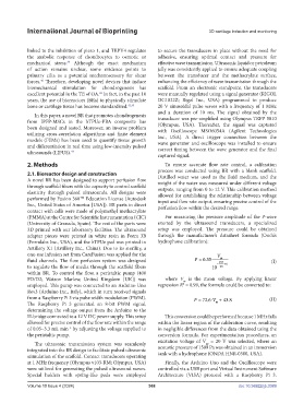
            (PMMA) at the Centre for Scientific Instrumentation (CIC)   For measuring the pressure amplitude of the P-wave
            (University of Granada, Spain). The rest of the parts were   exerted by the ultrasound transducers, a specialized
            3D printed with our laboratory facilities. The ultrasound   setup was employed. The pressure could be obtained
            adapter pieces were printed in white resin in Form 3B   through the manufacturer’s datasheet formula (Onda’s
            (Formlabs Inc., USA), and the bTPUe pad was printed in   hydrophone calibration):
            Artillery X1 (Artillery Inc., China). Due to its sterility, a
            one-use infusion set from CareFusion was applied for the                  V
                                                                                       m
            fluid channels. The flow perfusion system was designed           P = 035.  254                 (I)
            to regulate the flow of media through the scaffold fibers               10 −  20  +6
            within BR. To control the flow, a peristaltic pump (400
            FD/D2; Watson Marlow, United Kingdom [UK]) was        where  V  is the mean voltage. By applying linear
                                                                         m
            employed. This pump was connected to an Arduino Uno   regression R  = 0.99, the formula could be converted to:
                                                                         2
            Rev3 (Arduino Inc., Italy), which in turn received signals
            from a Raspberry Pi 3 via pulse width modulation (PWM).          P = 72 6.  V ⋅  + 438.        (II)
            The Raspberry Pi 3 generated an 8-bit PWM signal,                        m
            determining the voltage output from the Arduino to the
            H-bridge connected to a 12 V DC power supply. This setup   This conversion could be performed because 1 MHz falls
            allowed for precise control of the flow rate within the range   within the linear region of the calibration curve, resulting
            of 0.05–3.3 mL min  by adjusting the voltage supplied to   in negligible differences from the data obtained using the
                            −1
            the peristaltic pump.                              conversion formula. For experimentation procedures, an
                                                               excitation voltage of  V  = 20 V was selected, where an
               The ultrasonic transmission system was seamlessly                  pp
            integrated into the BR design to facilitate pulsed ultrasonic   acoustic pressure of 1509 Pa was obtained in an immersion
                                                               tank with a hydrophone (ONDA HNR-0500, USA).
            stimulation of the scaffold. Contact transducers operating
            at 1 MHz frequency (Olympus v103-RM; Olympus, USA)    Finally, the Arduino Uno and the Oscilloscope were
            were utilized for generating the pulsed ultrasound waves.   controlled via a USB port and Virtual Instrument Software
            Special holders with spring-like pads were employed   Architecture (VISA) protocol with a Raspberry Pi 3.

            Volume 10 Issue 4 (2024)                       368                                doi: 10.36922/ijb.3389

