
Page 386 - IJB-10-4
P. 386
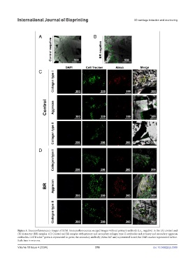
International Journal of Bioprinting                                   3D cartilage induction and monitoring


            Figure 5. Immunofluorescence images of ECM. Immunofluorescence merged images without primary antibody (i.e., negative) in the (A) control and
            (B) bioreactor (BR) samples. (C) Control and BR samples with primary and secondary collagen type II antibodies and primary and secondary aggrecan
            antibodies. Cell Tracker™ green is represented in green; the secondary antibody (Alexa 647 nm) is presented in red; the DAPI marker is presented in blue.
            Scale bars in microns.
            Volume 10 Issue 4 (2024)                       378                                doi: 10.36922/ijb.3389